Category: Exercise & Movement

Exercise has some of the strongest evidence of any intervention in Parkinson’s care. These guides cover aerobic, strength, balance, and Parkinson-specific programs like LSVT BIG and PWR! Moves, plus help for freezing of gait.

  • Stationary Cycling for Parkinson’s Patients: Exercise Program Alleviates Motor Dysfunction Naturally

    Stationary Cycling for Parkinson’s Patients: Exercise Program Alleviates Motor Dysfunction Naturally

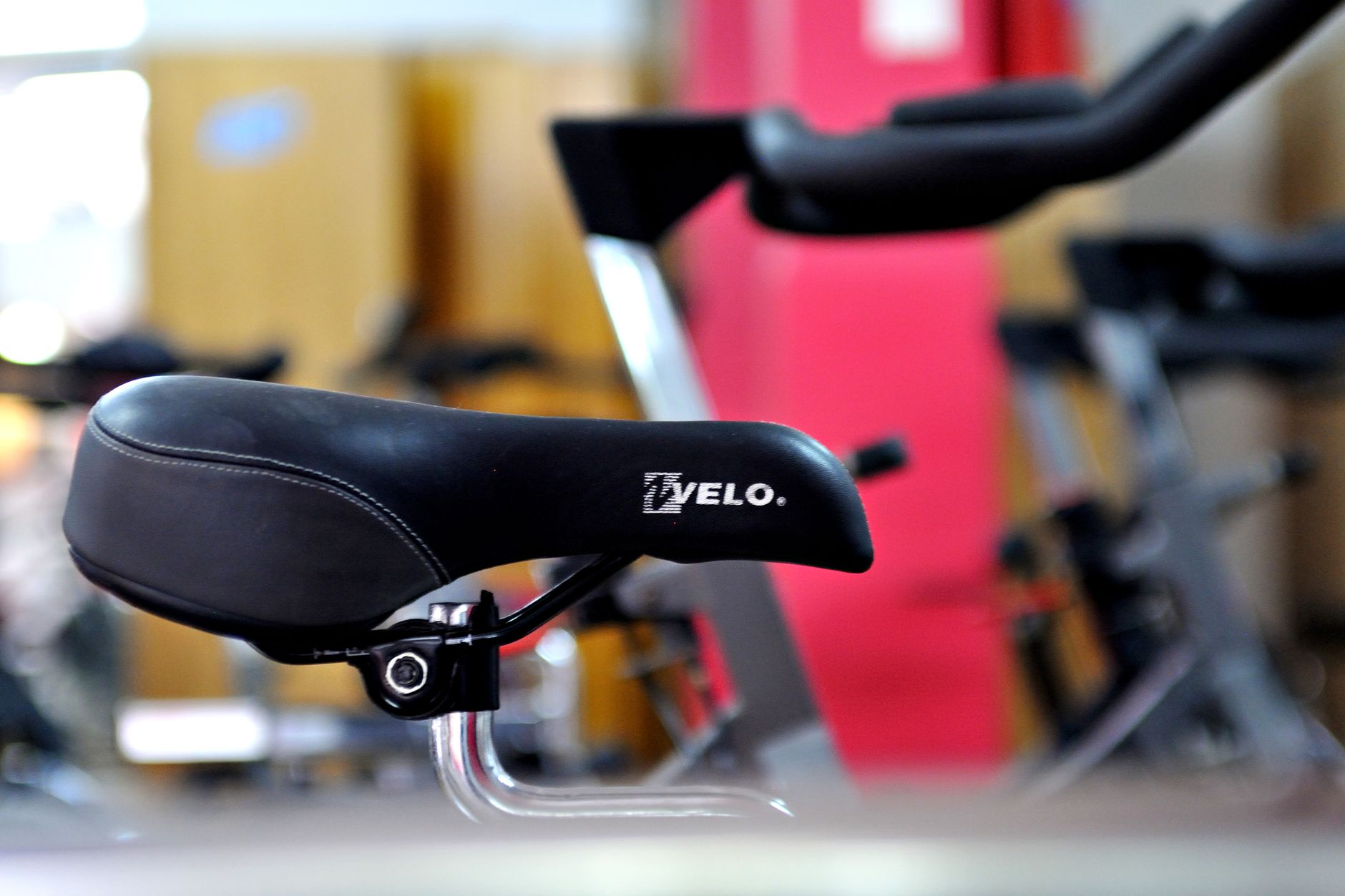
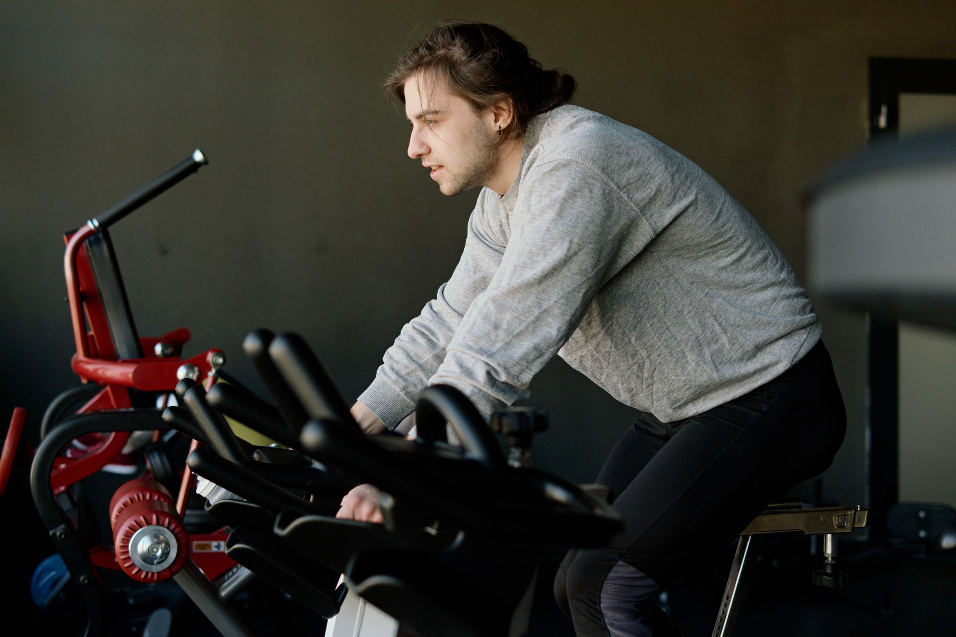
    Stationary cycling offers measurable relief from Parkinson’s motor symptoms through a form of exercise that engages large muscle groups while maintaining safety and control. Unlike walking, which can become restricted and unsteady as Parkinson’s progresses, cycling on a stationary bike forces continuous leg movement through a complete circular motion—a neurological pattern that appears to bypass some of the movement hesitation characteristic of the disease. A patient who struggled to initiate walking due to “freezing” episodes might find that pedaling comes more naturally, allowing them to exercise for extended periods without the cognitive burden of starting and stopping movements.

    The mechanism works partly through something called forced exercise—the bicycle pedal creates a pacing that the nervous system can follow, similar to how people with Parkinson’s walk more smoothly to the rhythm of music. Beyond the immediate physical benefits, regular stationary cycling activates neural pathways independent of the circuits damaged by Parkinson’s, potentially slowing decline in motor function over time. This is not a cure, but a sustained intervention that caregivers and patients increasingly use as part of comprehensive symptom management.

    Table of Contents

    How Does Stationary Cycling Specifically Help Parkinson’s Motor Symptoms?

    parkinson‘s disease damages dopamine-producing neurons, leaving people struggling with rigidity, tremor, bradykinesia (slow movement), and postural instability. Stationary cycling addresses these problems through forced, rhythmic movement that the body can execute without relying solely on the damaged dopamine system. The pedaling motion requires leg muscles to cycle through extension and flexion repeatedly, which keeps joints mobile and muscles engaged—both critical as Parkinson’s stiffness tends to worsen with inactivity.

    Studies have suggested that this type of continuous, paced exercise may enhance the recruitment of alternative motor pathways, allowing the brain to circumvent some of the movement deficits caused by neurodegeneration. The circular nature of pedaling is particularly advantageous because it eliminates the decision-making burden of discrete steps. In walking, each step requires the brain to initiate movement, decide pace, and manage balance—tasks that become cognitively exhausting for Parkinson’s patients. On a stationary bike, the patient pedals in a set motion that becomes almost automatic once initiated, reducing what clinicians call “cognitive load.” This can make longer exercise sessions feasible for people who tire quickly during conventional workouts, and it may explain why some patients report feeling less rigidity and moving more smoothly after cycling sessions.

    The Role of Forced Exercise and Neuroplasticity in Parkinson’s Management

    Forced exercise—meaning movement driven by an external mechanism like pedaling speed or a treadmill pace—activates different neural circuits than voluntary movement. When a Parkinson’s patient walks at their own pace, they rely heavily on the damaged striatum and other dopamine-dependent regions. But when pedaling a stationary bike with a set cadence, the rhythmic input bypasses some of this damaged circuitry by engaging cerebellar and motor cortex networks that remain relatively intact. Over weeks of consistent cycling, these alternative pathways may strengthen, potentially leading to measurable improvements in motor control even during non-cycling activities.

    However, there is an important limitation: the benefits of forced exercise appear to diminish if the cycling is performed passively or at very low intensity. A stationary bike that moves the patient’s legs without active participation—sometimes called passive cycling—shows minimal motor benefit in research. The patient must pedal with effort and maintain a steady cadence for the neuroplastic changes to occur. Additionally, stationary cycling benefits primarily the lower body; upper limb rigidity, tremor in the hands, or neck stiffness require separate interventions like resistance training or occupational therapy. Patients and caregivers sometimes overestimate how much cycling alone can improve overall motor function, leading to disappointment if upper body symptoms remain unchanged.

    Cardiovascular and Metabolic Benefits Beyond Motor Symptoms

    Parkinson’s disease often leads to reduced physical activity, sedentary behavior, and related cardiovascular deconditioning. stationary cycling, because it is low-impact and accessible to people with varying levels of motor impairment, allows patients to engage in aerobic exercise safely. Regular cycling strengthens the heart, improves circulation, and helps maintain a healthy body weight—all factors that reduce the risk of stroke, heart disease, and metabolic complications that can emerge as Parkinson’s progresses.

    The metabolic benefit extends to blood pressure regulation and blood sugar control. Sedentary Parkinson’s patients often develop insulin resistance and hypertension, both common complications of the disease. A patient who cycles for 30 minutes three times per week may not only move better but also stabilize blood sugar levels and reduce medication burden for hypertension. Additionally, maintaining aerobic fitness can improve energy levels and mood, both of which are often diminished in Parkinson’s disease independent of motor symptoms.

    Setting Up a Safe and Effective Stationary Cycling Program

    An effective stationary cycling program for Parkinson’s patients requires careful attention to bike setup and exercise parameters. The bike should be adjusted so the knee is slightly bent at the bottom of the pedal stroke—full leg extension creates stress on the knee joint, while excessive bending reduces the range of motion and muscular benefit. Seat height and handlebar position should be set to encourage upright posture without straining the back, as Parkinson’s patients often develop forward-stooping posture that can worsen with poor cycling ergonomics. Frequency and duration matter more than intensity for Parkinson’s motor improvement.

    Research has suggested that three sessions per week of 30 to 45 minutes at moderate intensity produces measurable motor benefits over 12 to 24 weeks. This is substantially more accessible to most patients than the high-intensity protocols sometimes promoted for general fitness, and it aligns with what people with Parkinson’s can realistically sustain without exhaustion or injury. A patient who cycles twice per week for 20 minutes is likely to gain cardiovascular benefit but may miss the neuroplastic motor improvements that come with more consistent, higher-volume training. The trade-off is between convenience and effectiveness; caregivers must help negotiate this balance based on the patient’s energy levels, disease stage, and schedule.

    Monitoring Progress and Recognizing When Cycling Alone Is Insufficient

    As Parkinson’s progresses, the motor benefits of stationary cycling may plateau or diminish, particularly if the disease is advancing rapidly or if medication dosing is suboptimal. A patient who experienced marked improvement in the first 6 months might find that benefits stall or that symptoms return to baseline by month 12. This does not mean cycling is failing; rather, it reflects the progressive nature of the underlying neurodegeneration. Continuation of cycling is still warranted for cardiovascular health and to potentially slow further decline, but expectations should be adjusted realistically.

    One warning: some patients experience increased tremor, stiffness, or dyskinesia (involuntary writhing movements) when cycling at certain intensities or times of day. If tremor worsens during pedaling, it may indicate that the cycling cadence is too fast or that the patient is cycling during a time of day when medication effectiveness is waning. Adjusting cadence downward or shifting the cycling time to peak medication hours (typically 30 to 60 minutes after taking a dose) can often resolve this. Patients should never force themselves through worsening symptoms; instead, they should communicate changes to their neurologist or therapist to optimize both medication and exercise timing.

    Combining Stationary Cycling with Physical and Occupational Therapy

    Stationary cycling works best as part of a broader exercise regimen that includes balance training, strength work, and flexibility exercises. A patient who cycles three times per week but does not perform balance exercises remains at high risk for falls—cycling does not improve balance or proprioception, the senses that tell the body where it is in space. Adding a second or third exercise modality, such as tai chi, resistance training with a physical therapist, or gait training, creates a more comprehensive motor intervention.

    For example, a patient might cycle on Mondays and Fridays, attend a physical therapy session on Wednesday focused on balance and gait, and practice home exercises on alternate days. Occupational therapists can also help patients perform upper-body and fine motor activities that cycling does not address. Hand exercises, writing practice, or resistance work with the arms and shoulders target the tremor and rigidity in the upper body that often most affects quality of life. The combination approach takes more time and coordination but delivers better overall motor and functional outcomes than cycling alone.

    Long-Term Sustainability and the Role of Caregiver Support

    One of the strongest predictors of long-term adherence to a stationary cycling program is consistent caregiver support and encouragement. Parkinson’s fatigue and depression—both common—can make it tempting to skip exercise sessions, especially when the cognitive effort of motivating oneself is high.

    A caregiver who schedules the cycling time, helps the patient get to the bike, and provides positive reinforcement dramatically increases the likelihood that the patient will sustain the program over months and years. Some patients benefit from cycling while listening to music or audiobooks, a strategy that provides dual cognitive stimulation and makes sessions feel less monotonous. Others find group cycling classes designed for Parkinson’s patients particularly motivating; these specialized programs, increasingly available at hospitals and community centers, offer both the forced-exercise benefit and the social engagement that can lift mood and reinforce commitment.

    You Might Also Like

  • Stationary cycling proven to ease Parkinson’s movement complications in seniors

    Stationary cycling proven to ease Parkinson’s movement complications in seniors

    Stationary cycling has emerged as a practical exercise intervention that can help reduce motor complications in people with Parkinson’s disease, particularly in older adults who face mobility challenges. Research suggests that regular cycling on a stationary bike addresses several movement problems central to Parkinson’s—including stiffness, balance difficulties, and the shuffling gait that characterizes the disease. A person with Parkinson’s who cycles at moderate intensity several times per week may notice gradual improvements in walking smoothness and an ability to turn more naturally, as the repetitive pedaling motion seems to engage motor systems in ways that combat the progressive neurological changes of the disease.

    The mechanism appears to involve both the physical conditioning benefits of steady-state aerobic exercise and something more specific to cycling’s rhythmic, bilateral leg movements. Unlike some other exercises that require complex coordination or high impact on joints, stationary cycling allows people with reduced mobility to maintain cardiovascular fitness and motor function while sitting safely and supported. This makes it accessible even to those with significant tremor, rigidity, or postural instability—concerns that might prevent participation in walking programs or gym classes.

    Table of Contents

    How Does Stationary Cycling Address Parkinson’s Movement Difficulties?

    parkinson‘s disease affects the basal ganglia, brain regions that coordinate smooth, automatic movement. People with the condition often experience bradykinesia (slow movement), rigidity, tremor, and loss of the automatic quality of everyday actions like walking or swinging the arms. Stationary cycling may counteract these problems by providing rhythmic external cues—the steady cadence of pedaling—that the brain can use to organize movement. This is similar to how listening to music with a steady beat can help some people with Parkinson’s walk more fluidly; the external rhythm bypasses some of the damaged circuits and allows movement to proceed more naturally.

    The aerobic benefits of cycling also matter. Regular cardiovascular exercise increases blood flow to the brain and may promote the growth of new neural connections, potentially slowing cognitive decline in Parkinson’s. Additionally, the leg muscles engaged during cycling are among the largest in the body, so cycling provides substantial cardiovascular stimulus without the impact stress of running. Someone who was previously unable to walk for long distances might cycle for 20 or 30 minutes, building stamina and lower-body strength in a way that translates to better walking ability and reduced fatigue in daily life.

    The Specific Benefits and Real Limitations of Stationary Cycling

    Stationary cycling offers several advantages over treadmill walking or outdoor cycling for older adults with Parkinson’s. The bike is stationary, eliminating fall risk from balance loss or sudden freezing episodes. The seat provides support, and the pedals are confined to a predictable path, so tremor or involuntary movements are less likely to derail the exercise. Many people find they can maintain cycling for longer periods than they could manage other forms of exercise, which means more total movement and greater conditioning gains. However, important limitations exist.

    Stationary cycling does not address some Parkinson’s motor problems equally well. Postural instability and balance deficits, for instance, may actually worsen if cycling is the only exercise, because balance is not challenged. A person who becomes stronger on the bike but does not work on balance and core stability may still be at high risk of falling during everyday activities like reaching for items or turning while standing. Additionally, cycling does not engage the upper body, arms, and trunk in ways that some other exercises do. medication timing also matters—symptoms fluctuate as Parkinson’s medications wear on and off, and cycling during “off” periods when symptoms are most severe may be ineffective or unsafe, so the best time to cycle is often when medication is working well.

    How Intensity and Frequency Shape Outcomes

    research on exercise for Parkinson’s suggests that consistency and moderate intensity matter more than occasional high-effort sessions. Cycling three to four times per week, for 30 to 45 minutes per session, at a pace that feels moderately challenging—roughly 50 to 70 percent of maximum heart rate—appears to produce measurable improvements in gait speed and stride length within weeks to months. A person might start with two 20-minute sessions per week and gradually build to longer, more frequent workouts as conditioning improves.

    One complicating factor is medication response. Someone taking levodopa or other Parkinson’s medications might notice that benefits of cycling are most evident during windows when medication is providing good symptom control. If cycling happens during an “off” period when the medication is wearing off, motivation and performance drop, and the neurological stimulus may not be as strong. Conversely, cycling consistently during “on” times—such as an hour after taking medication—can reinforce better movement patterns and may lead to longer-lasting improvements even during off periods.

    Comparing Stationary Cycling to Other Parkinson’s Exercises

    Stationary cycling is often compared to treadmill walking, resistance training, and tai chi in discussions of Parkinson’s exercise. Treadmill walking can help with gait and provides more direct training for the movement pattern people need in daily life, but treadmills carry fall risk for those with balance problems and require careful supervision. Resistance training (weight lifting or elastic band exercises) is excellent for addressing muscle weakness but does not provide the same cardiovascular benefits or the rhythmic motor engagement that cycling offers. Tai chi is valuable for balance and can improve postural control, but it requires coordination and concentration that some people with advanced Parkinson’s find challenging.

    A practical approach for many people involves combining stationary cycling with other exercises. Someone might cycle three times per week for conditioning and rhythm, do resistance exercises twice per week to maintain muscle and bone density, and practice balance or tai chi on separate days to address postural instability. This combination addresses more aspects of Parkinson’s movement dysfunction than cycling alone could achieve, though it requires more time and planning. The tradeoff is that a broader exercise program is more sustainable long-term because it addresses multiple concerns and reduces the monotony of doing the same activity repeatedly.

    Medication Interactions and Safety Concerns

    People taking Parkinson’s medications need to be aware that exercise can affect symptom severity and medication needs over time. As fitness improves through regular cycling, some individuals find that their medications feel more effective or that they need less frequent dosing adjustments. This is encouraging but also requires communication with the treating neurologist, as medication prescriptions may need adjustment if exercise-related changes alter symptom patterns. Never adjust medication timing or dose based solely on feeling better during or after exercise.

    A significant safety concern is heat and dehydration. Stationary cycling indoors can lead to sweating and fluid loss, especially if the room is warm or if someone is not accustomed to exercise. Dehydration can worsen Parkinson’s symptoms, including tremor and rigidity, and can also affect medication absorption. People cycling should drink water before, during, and after the session—a simple step that many overlook. Additionally, some Parkinson’s medications can affect blood pressure regulation, and exercise raises blood pressure temporarily; older adults with Parkinson’s should monitor for dizziness or unusual heart palpitations during or after cycling and should speak with their doctor if these occur.

    Setting Up a Home Cycling Program

    Many people with Parkinson’s find stationary cycling most sustainable when the bike is in a convenient, visible location at home, preferably where they can watch television or listen to music during the workout. This removes barriers related to getting to a gym or managing transportation on days when symptoms are difficult.

    Recumbent stationary bikes—where the rider sits back and extends legs forward rather than sitting upright—are often easier for people with balance concerns or spinal stiffness, as they provide more support and do not require the same postural control as upright bikes. A practical setup might include a stationary bike in a living room, a mirror to allow monitoring of posture during cycling, and perhaps a small table nearby for water and a towel. Many people benefit from setting a consistent time for cycling—such as one hour after taking medication in the morning—to build a habit and ensure it happens regularly rather than sporadically.

    Long-Term Outlook and Individual Variation

    Improvements from stationary cycling in Parkinson’s are typically gradual and may plateau after several months, rather than showing continuous dramatic gains. Someone might notice clearer changes in walking smoothness or reduced stiffness over the first two to three months, then find that further gains come more slowly. This does not mean cycling has stopped working; rather, the exercise may be maintaining function and slowing decline rather than producing visible new improvements.

    Over longer periods—years—consistent cycling appears to help preserve mobility and independence in ways that matter for quality of life, even if the changes are subtle month to month. Individual responses vary considerably depending on Parkinson’s stage, age, other health conditions, and medication regimen. A person in early Parkinson’s might see dramatic gait improvements from cycling, while someone in mid-to-late stage disease might use cycling primarily to maintain strength and cardiovascular fitness rather than expecting major symptom reversal. There is no single “right” cycling program that works for everyone, so working with a physical therapist or neurologist to tailor cycling intensity, duration, and frequency to individual abilities and goals increases the likelihood of sustained benefit.


    You Might Also Like

  • Parkinson’s movement monitoring: New sensor technology enables at-home tracking

    Parkinson’s movement monitoring: New sensor technology enables at-home tracking

    New wearable sensor technology has created practical opportunities for Parkinson’s disease patients to track their own movement patterns at home, without requiring frequent trips to the clinic. These devices use accelerometers and gyroscopes—the same motion-sensing components found in smartphones—to detect tremors, measure walking speed, assess arm swing, and quantify rigidity or slowness of movement. A patient might wear a small sensor on the wrist or ankle and receive daily reports showing how their symptoms fluctuate, which symptoms are most prominent at different times, and how their medication appears to be working throughout the day.

    For people living with Parkinson’s, this continuous monitoring addresses a real clinical gap. Neurologists typically see patients every three to six months, observing only a snapshot of behavior during a clinic visit. Between appointments, patients and their caregivers are left estimating whether symptoms are stable, worsening, or responding well to treatment changes. At-home sensor monitoring provides a more complete picture of movement patterns across the real world, with data collected over weeks or months rather than minutes spent in an examination room.

    Table of Contents

    How Wearable Sensors Detect Parkinson’s Movement Changes

    Wearable accelerometers measure motion along multiple axes, detecting the characteristic tremor frequencies that distinguish Parkinson’s tremor from other types of shaking. Gyroscopes add rotational data, capturing the subtler jerking or oscillating movements that may not show up on simple motion sensors. When a patient wears a sensor on the wrist or forearm, for instance, the device continuously records acceleration and rotation, then processes that data through software algorithms that recognize patterns associated with Parkinson’s symptoms. Bradykinesia—the slowing of movement that defines Parkinson’s disease—shows up in sensor data as reduced acceleration during functional tasks like reaching, turning a doorknob, or getting out of a chair.

    Gait analysis is equally revealing: sensors can measure stride length, walking speed, the regularity of steps, and the degree to which arm swing is reduced, all of which are hallmarks of Parkinson’s locomotion. Walking tests done in clinics last seconds; home sensors record actual behavior over entire days, capturing how symptoms change with fatigue, time of day, medication timing, and activity level. The advantage over clinical observation is that sensors eliminate observer bias and provide precise, objective measurements. A neurologist watching a patient walk down a hallway can describe “slow walking” or “reduced arm swing,” but a sensor provides numbers: stride length reduced by 15 percent, walking speed at 3.2 kilometers per hour instead of a baseline of 4.1. This specificity helps clinicians spot medication effectiveness more clearly and detect subtle changes that might otherwise be missed.

    Sensor Placement and Which Symptoms Are Trackable

    Most research and commercial systems use wearable sensors on the wrist, ankle, lower back, or a combination of these locations. Wrist-worn devices are convenient and address the tremor and arm movement components of Parkinson’s. Ankle or foot sensors capture gait, detecting freezing of gait—the sudden inability to initiate or continue walking—which is a common and dangerous symptom. Back-mounted sensors measure postural sway and changes in body position. Depending on where the sensor sits, different aspects of the disease are visible. However, not all Parkinson’s symptoms are equally trackable by sensors.

    Tremor and slowness of movement translate reliably into sensor data. But rigidity—the muscle stiffness that resists passive movement—is harder for an external sensor to capture, because rigidity is felt during a clinical examination but may not produce distinctive motion patterns during daily activity. Cognitive symptoms like slow thinking or memory problems are entirely invisible to movement sensors. This means that sensor monitoring is best viewed as one input into overall symptom management, not a complete diagnostic or monitoring system. A limitation that often surprises patients is that wearing the device itself can influence movement patterns. The presence of a sensor on the wrist can make some people hold their arm differently or walk more carefully, knowing they are being monitored. Data from carefully controlled laboratory tests doesn’t always match data from free-living situations, even when the sensor is measuring the same underlying movement.

    How Patients Use Daily Movement Data to Manage Symptoms

    The central value of at-home sensor monitoring is showing patients and caregivers how their movement varies throughout the day and across days. A patient might notice that tremor is worst in the morning before medication takes effect, moderate by midday, and much better by evening—information that might support a conversation with their neurologist about timing or dosing. Someone with Parkinson’s might discover that their walking speed drops noticeably in late afternoon, perhaps due to medication wearing off, or that certain activities consistently trigger freezing episodes. Caregivers also benefit from objective movement data.

    A spouse or adult child concerned that symptoms are worsening has concrete evidence to share with the physician, rather than relying on memory or general impression. This is particularly useful when the person with Parkinson’s has cognitive changes that make self-reporting less reliable. If a caregiver notices deterioration in the sensor data over weeks, they can bring that trend to a medical appointment rather than waiting for a crisis or a sudden worsening the doctor observes in the clinic. Patients sometimes use the data to adjust their own routines: taking medication before anticipated activities, avoiding situations where gait freezing is likely to occur, or scheduling physically demanding tasks when their sensor data shows movement is typically best.

    Cost, Insurance, and Access Realities

    Wearable sensor systems for Parkinson’s monitoring range widely in price, from lower-cost consumer fitness trackers adapted for symptom tracking to specialized medical-grade devices costing several hundred dollars per unit or more. Setup typically involves downloading an app, pairing the sensor device via Bluetooth, and allowing the system to begin collecting and storing data. Most systems transmit data to a cloud platform where algorithms process it and generate reports. Insurance coverage for these devices remains limited and varies by region and plan. Some systems are cleared by regulatory agencies like the FDA for clinical use, which increases the likelihood of insurance reimbursement, but reimbursement is not guaranteed.

    Out-of-pocket costs can be a barrier, particularly for patients on fixed incomes or those without strong private insurance. This creates an equity issue: patients with resources can access detailed movement monitoring, while others cannot, even though the technology might be equally beneficial. The tradeoff is between cost and convenience. A patient who can afford ongoing device use gets continuous data and the ability to work with their neurologist on subtle medication adjustments. A patient without access might see their neurologist only every six months, missing opportunities to intervene if symptoms are changing. Neurologists and patients should weigh whether the cost is justified by the specific clinical questions they are trying to answer—for instance, fine-tuning a medication dose over weeks—versus whether less frequent in-clinic assessments would be sufficient.

    Accuracy Challenges and Data Interpretation Limitations

    Wearable sensor accuracy for Parkinson’s is better than nothing but not perfect. Different algorithms interpret the same motion data differently, and the software that translates raw accelerometer readings into clinical measurements like “tremor severity” or “walking speed” varies between manufacturers and research teams. A patient using one system might receive a report that their tremor is moderately improved, while the same movement data processed by a different algorithm might show minimal change. Environmental factors also affect sensor readings. A patient tremoring while sitting in a car, while riding a bus, or while feeling anxious produces different sensor data than the same tremor while standing still. Temperature can affect accelerometer accuracy.

    Sweat or moisture can interfere with some devices. These real-world variations mean that comparing a patient’s data across weeks or months requires some caution—changes might reflect actual symptom progression, or they might reflect differences in how the patient was moving when the sensor was recording. A critical limitation is that patients and families often misinterpret what the data means. A decrease in measured arm swing might indicate worsening Parkinson’s, or it might mean the patient wore the sensor slightly differently that day, or held their arm in a different position during their morning walk. Without training, patients can become anxious over normal day-to-day variation or falsely reassured by data artifacts that don’t reflect true symptom change. This is why sensor data is most useful when interpreted with guidance from a clinician familiar with both Parkinson’s disease and the specific system being used.

    Data Storage and Privacy Considerations

    Most sensor systems store data on manufacturers’ cloud platforms, allowing patients and providers to access reports from any device with internet connection. This convenience comes with privacy considerations. Medical data involving movement and symptom tracking is sensitive health information.

    Patients should understand what company owns their data, how long it is retained, whether it might be used for research, and what protections exist if the company is breached or changes ownership. Some patients prefer standalone systems that record data locally on the device itself, with optional uploads to a personal account, rather than automatic cloud transmission. Others find the convenience of cloud data storage outweighs privacy concerns. Neither approach is inherently right; the choice depends on individual comfort with data sharing and how critical the reporting features are to their symptom management.

    Patient Adoption and Long-Term Use in Real Life

    Early experience with patient-deployed wearable sensors shows that people do use them—some devices report that patients wear them and sync data regularly for months—but adoption isn’t universal. Some patients abandon the devices after weeks because they find the daily reports overwhelming or anxiety-inducing, because wearing the sensor becomes uncomfortable or inconvenient, or because the data doesn’t feel actionable.

    Others find the technology genuinely helpful for collaborating with their neurologist. Clinical practices that have integrated sensor monitoring typically find it most valuable when the device is used for a specific, time-limited question—”Let’s track tremor severity for the next month to see if this medication change is helping” rather than as an indefinite, open-ended monitoring project. This focused approach gives patients a clear purpose for wearing the sensor and neurologists a concrete reason to interpret the data, making the effort feel worthwhile rather than like surveillance.

    Frequently Asked Questions

    Can wearable sensors detect all Parkinson’s symptoms?

    Wearable sensors effectively track tremor, slowness of movement, and gait changes. They cannot reliably measure rigidity or detect cognitive or emotional symptoms. Sensor monitoring works best as one tool alongside clinical assessments, not as a complete replacement.

    Will my insurance cover a Parkinson’s monitoring device?

    Coverage varies by insurance plan and device type. Devices cleared for clinical use by regulatory agencies have a better chance of reimbursement, but coverage is not guaranteed. Contact your insurance provider and ask whether your neurologist can request coverage for your specific device.

    What if the data seems wrong or contradicts how I feel?

    Day-to-day variations in movement, sensor wear, and how you hold the device can all affect readings. Don’t assume a single day’s data shows a real change in your condition. Discuss trends over weeks or months with your neurologist, and mention if you notice the data doesn’t match how you actually feel.

    Is my movement data private?

    Most systems store data on company cloud servers. Review the manufacturer’s privacy policy, understand how long your data is kept, and whether it might be used for research. Some systems allow local data storage if privacy is a primary concern.

    When should I consider adding a wearable sensor to my Parkinson’s care?

    Sensor monitoring is most useful when you and your neurologist have a specific question to answer, such as testing whether a medication change is working or tracking symptom patterns that aren’t clear from clinic visits. Open-ended monitoring without a defined goal is less likely to sustain patient engagement.


    You Might Also Like

  • How Boxing Training Helps Parkinson’s Patients Improve Movement Control

    How Boxing Training Helps Parkinson’s Patients Improve Movement Control

    Boxing training helps Parkinson’s patients improve movement control by engaging multiple neural pathways that bypass or strengthen the areas of the brain affected by the disease. The combination of rhythmic footwork, directional punch combinations, and visual focus creates a framework for movement that the brain can follow and execute more consistently than unstructured daily activities. A Parkinson’s patient who struggles to initiate a step forward on command may find they can move fluidly when executing a boxing combination—the repetition, timing, and external cues act as a neurological workaround that actually enhances motor function over time.

    Boxing differs from traditional exercise in that it demands simultaneous attention to multiple movement components: weight transfer, limb coordination, visual tracking, and timing. For Parkinson’s patients, who often experience bradykinesia (slowness of movement), rigidity, and difficulty initiating motion, these structured challenges stimulate adaptive changes in how the remaining motor circuits function. The programs that have gained traction, pioneered through initiatives like Rock Steady Boxing, focus on non-contact boxing drills rather than sparring, making the activity accessible to people across the severity spectrum of Parkinson’s disease.

    Table of Contents

    Why Rhythmic Movement and External Cueing Help Parkinson’s Patients Control Motion

    parkinson‘s disease primarily affects the basal ganglia and substantia nigra, regions that coordinate automatic and smooth movement. Without these neural systems functioning optimally, patients must rely more heavily on conscious, deliberate motor control—which requires engagement of other brain regions like the motor cortex and cerebellum. Rhythmic external cues (like music, a coach’s verbal count, or the visual target of a punching bag) help bypass the damaged circuits by providing external structure that the intact motor systems can lock onto and follow. When a Parkinson’s patient attempts an everyday movement like walking without external cuing, the motor command becomes fragmented and slow. But introduce a metronome, a marching band, or in the case of boxing, a rhythmic drill sequence, and the same patient can move with dramatically greater speed and fluidity.

    This phenomenon, called external cueing, is well-documented: patients who walk to music move faster and with better gait quality than when walking in silence. Boxing training essentially embeds this cueing principle into a structured physical activity, providing both rhythmic auditory input and visual targets that guide movement. The cerebellum, which plays a major role in timing and coordination, becomes more engaged during boxing training than during typical aerobic exercise. Because boxing demands precise timing between upper and lower body movements, the cerebellum must continuously adjust and refine motor output. Over weeks and months of consistent training, this repeated activation appears to strengthen the cerebellar contribution to movement control, potentially compensating for some of the basal ganglia dysfunction that Parkinson’s causes.

    Improvements in Balance, Speed, and Movement Initiation

    One of the most disabling symptoms of Parkinson’s disease is postural instability—the loss of automatic balance corrections. A patient who stumbles cannot quickly catch themselves because the reflex arc that normally coordinates limb movement is impaired. Boxing training, by demanding rapid weight shifts and stance changes, challenges and gradually retrains these balance mechanisms. A boxer must shift weight from one foot to the other in sequence, maintain an athletic stance, and control the transition between different positions—all skills that directly address postural control deficits. Bradykinesia, or slowness of movement, improves measurably with boxing training in part because the activity demands speed. A patient does not move slowly because their muscles are weak; they move slowly because the motor signal itself is diminished and delayed.

    When a boxing coach cues a patient to throw a combination at a brisk tempo, the patient must overcome that internal resistance to speed and execute the movement faster than they would naturally initiate. This repeated practice at higher speeds appears to reset the patient’s internal movement tempo, and improvements often carry over to non-boxing activities. However, not all Parkinson’s patients benefit equally, and progression of the disease can reduce the gains. A patient in early to mid-stage Parkinson’s may see marked improvements in walking speed and balance within a few weeks of boxing training, while a patient with advanced disease and significant cognitive decline may struggle to follow multi-step boxing combinations. Additionally, the benefits appear to require ongoing practice; patients who stop boxing often experience gradual erosion of the gains within weeks to months. This means boxing is not a one-time intervention but rather a long-term commitment similar to physical therapy.

    Motor Learning and Neuroplasticity in Parkinson’s Training

    The brain retains its ability to form new movement patterns and strengthen neural connections throughout life, a property called neuroplasticity. Parkinson’s disease does not erase this capacity; it changes the ease with which new motor learning occurs. Boxing training exploits neuroplasticity by repeatedly practicing complex, goal-directed movements in a structured environment. Each repetition reinforces the motor pattern, and the involvement of the cerebellum, motor cortex, and prefrontal cortex in learning creates redundancy—if one pathway is partially degraded, the others can partially compensate. Unlike passive movement (such as having limbs moved by another person) or single-plane repetitive motion (like using a treadmill), boxing requires the patient to actively solve the motor problem of how to execute a specific combination.

    This active, problem-solving quality appears to enhance learning. A patient who mechanically throws a punch because they are told to does so with less neural engagement than a patient who is learning a new combination and actively correcting their technique. Over time, this active learning process seems to establish more durable and transferable motor improvements. Interestingly, some of the neuroplastic changes appear to involve regions that are not directly part of the motor system, including areas involved in attention and executive function. Because boxing demands mental focus and decision-making (which combination next, how fast, where is the target), the prefrontal cortex and parietal regions are engaged. This cognitive engagement may contribute to improvements in non-motor symptoms as well, including mood and executive function.

    Practical Considerations for Starting a Boxing Program

    A Parkinson’s patient interested in boxing training should work with a coach or physical therapist experienced with the disease, as standard boxing instruction is not designed for the motor deficits Parkinson’s patients face. Specialized programs adapt boxing drills to accommodate tremor, rigidity, and balance problems. For example, instead of rapid footwork patterns, a patient might begin with simple stance work and slow, controlled punches, progressing over weeks to faster combinations as control improves. The training schedule matters significantly. Research on structured boxing programs suggests that three sessions per week, each lasting 45 minutes to an hour, produces noticeable improvements within 4 to 8 weeks. A single weekly session may provide maintenance benefits but appears less effective for generating improvement.

    This frequency requirement is more demanding than many traditional Parkinson’s exercise programs, and adherence is often the limiting factor. Patients who are highly motivated or who train in a group setting tend to stick with boxing longer than those who attempt it alone. The progression of training also requires care. A boxing program that is too simple may not challenge the motor system sufficiently to drive neuroplastic change; one that is too difficult can discourage the patient or increase fall risk. A good coach adjusts complexity and speed dynamically, pushing the patient to work hard without crossing into unsafe territory. The non-contact nature of these programs is important; a Parkinson’s patient should never attempt to spar or exchange blows, as slowed reactions and balance problems make contact boxing unsafe.

    Safety Concerns and Limitations in Boxing for Parkinson’s Patients

    Because Parkinson’s patients often have postural instability and may experience sudden freezing episodes (temporary inability to move), boxing training carries fall risk. A patient who suddenly freezes mid-combination or who loses balance while throwing a punch could fall and injure themselves. For this reason, boxing programs for Parkinson’s patients must include environmental modifications such as training near a wall or rail, wearing appropriate footwear, and having a spotter or coach present. Patients with advanced balance deficits or a history of falls should discuss boxing with their neurologist before starting and consider more conservative forms of exercise if the neurologist advises against it. Not all motor symptoms improve equally with boxing. Tremor, in particular, may or may not respond to boxing training. Some patients experience reduced tremor with activity, but the effect is often temporary and limited to the moving limb.

    Patients whose Parkinson’s disease includes significant dystonia (muscle rigidity and twisting) may find that intense exercise, paradoxically, aggravates symptoms. Medication timing also matters; a patient whose medication is wearing off may move so stiffly that boxing becomes impossible or risky. Optimal training typically occurs during the patient’s window of best medication effect. Cognitive decline is another boundary condition. Parkinson’s disease is progressive, and many patients eventually develop cognitive symptoms ranging from mild memory problems to dementia. A patient who cannot remember a three-punch combination or who becomes confused during a session will not benefit as much from boxing training and may become frustrated. As the disease progresses, patients may need to transition to simpler, less cognitively demanding forms of exercise. Boxing is not universally appropriate for all stages of Parkinson’s disease.

    Group Training and Psychological Benefits

    One of the most consistent observations from Parkinson’s boxing programs is that group training produces better adherence and higher reported satisfaction than individual coaching. Training in a class with other Parkinson’s patients creates social connection, accountability, and a shared sense of purpose. Patients often report that they feel less isolated and more motivated when surrounded by others facing the same disease. The psychological boost—reduced depression, increased confidence, a sense of agency—may be as important as the motor improvements for long-term wellbeing.

    Group classes also allow for peer learning and encouragement. When a patient sees another person with similar symptoms progress from barely throwing a punch to executing combinations with control, the motivation to persist increases. The instructor can modify movements for different ability levels within the same class, allowing experienced patients to work harder while newer patients learn fundamentals. This heterogeneity of a good group class makes it more effective than a one-size-fits-all program.

    Long-Term Maintenance and Integration Into Parkinson’s Care

    The improvements from boxing training are not permanent if the training stops. Patients who discontinue boxing gradually lose gains in speed and balance control, typically over a period of weeks to months depending on disease stage and individual variability. For this reason, long-term improvement in Parkinson’s requires boxing to become an ongoing part of the patient’s routine, integrated alongside medication, physical therapy, and other disease management strategies. Some patients benefit from continuing at a reduced frequency (one to two sessions per week) once they reach a plateau, which maintains benefits with lower time and logistical burden.

    Boxing training works synergistically with medication and other therapies. A patient who trains regularly and manages their medication schedule often achieves better overall motor control than either intervention alone. Because boxing activates multiple neural systems and uses external cueing, it engages brain mechanisms that are not fully engaged by medication alone. For a comprehensive Parkinson’s management plan, boxing can serve as a core physical strategy, occupying a role similar to but more intensive than conventional physical therapy. The specific advantage of boxing is that it demands continuous attention and adjustment, which may provide greater neuroplastic stimulus than more rote exercises.


    You Might Also Like

  • Community Dance Programs Help Parkinson’s Patients Regain Movement

    Community Dance Programs Help Parkinson’s Patients Regain Movement

    Community dance programs offer Parkinson’s patients a way to work on movement, balance, and coordination in a social setting that feels more like recreation than therapy. Unlike traditional exercise classes, dance engages the brain’s rhythmic and motor systems simultaneously, potentially helping people with Parkinson’s move more fluidly and with greater confidence. These programs range from specialized classes designed specifically for Parkinson’s to mainstream dance studios that welcome participants of all abilities, creating environments where people can pursue movement on their own terms rather than in isolation.

    Dance addresses several core challenges in Parkinson’s disease. Many patients experience slowness of movement, stiffness, difficulty initiating steps, and reduced balance—all issues that dance practice can target. The combination of music, structured movement patterns, and peer interaction creates a multisensory experience that engages different brain pathways than medication alone, sometimes producing improvements that extend beyond the class itself.

    Table of Contents

    How Dance Engages Movement Pathways in Parkinson’s Disease

    Dance works differently in the Parkinson’s brain than conventional exercise. The rhythmic, predictable nature of music and choreography can bypass some of the motor-control difficulties that characterize the disease. When people with Parkinson’s move to external rhythm—a beat, a musical phrase, or a partner’s movement—they often move more smoothly and with less hesitation than when initiating movement on their own. This phenomenon, called rhythmic cueing, is one reason dance can be especially helpful where other activities might not be. The cognitive engagement is equally important.

    Dance requires attention to music, to other dancers, to your own body position, and to the choreography itself. This multitasking demand activates multiple brain regions, potentially strengthening neural networks affected by Parkinson’s. A person who struggles to walk in a straight line on their own may find they can navigate a dance pattern more successfully because their brain is focused outward rather than inward on the effort of movement. Balance and postural control improve through dance practice in ways that matter for daily life. Dance classes emphasize weight shifting, turning, and maintaining posture while moving—exactly the skills that deteriorate in Parkinson’s and that lead to falls. Regular practice in a supported environment means patients practice these risky movements repeatedly, building confidence and muscle memory.

    Finding and Evaluating Community Dance Programs

    Not all dance programs are equally suitable for Parkinson’s patients. Some classes are designed specifically for the disease, with instructors trained in Parkinson’s motor symptoms and how to modify movements appropriately. Others are mainstream classes—ballet, contemporary, salsa—that welcome people with various abilities but lack specialized adaptation. There are trade-offs: specialized Parkinson’s classes often have smaller groups and instructors who understand fatigue patterns and medication timing, but they may be less available geographically. Mainstream classes offer more variety and social integration with non-Parkinson’s dancers, but require self-advocacy and may lack appropriate modifications. Cost and accessibility are real barriers.

    Specialized Parkinson’s dance programs are sometimes subsidized by nonprofits or offered through hospitals and research centers, making them more affordable. Others operate independently and may be expensive. Transportation can be another obstacle; some programs offer on-site parking or partner with local medical centers that are easier to reach than neighborhood dance studios. Quality matters significantly. An instructor who understands Parkinson’s—how rigidity affects flexibility, how freezing episodes might occur, how medication timing influences performance—can make the difference between a beneficial class and a frustrating one. Programs that allow observers or offer trial classes let patients and caregivers assess whether the instruction, pacing, and social environment are suitable before committing.

    The Social and Psychological Dimensions of Dance

    Community dance is not only about the neurological benefits of movement. The social connection is powerful. Parkinson’s can be isolating; people may withdraw from activities as symptoms progress, or feel self-conscious about their movement. A dance class provides structured peer interaction, a sense of shared purpose, and an identity beyond illness.

    Dancers support each other, celebrate improvements, and sometimes continue friendships outside the class. The psychological shift can be significant. Rather than thinking of movement as a symptom problem to be managed, dancers think of themselves as artists or athletes pursuing a skill. This reframing—from “I can’t move normally” to “I am learning to dance”—affects how people relate to their bodies and their disease. Some participants report that the confidence gained in dance carries over to other daily activities.

    Practical Considerations for Getting Started

    Before joining a program, patients should consult their neurologist or movement disorder specialist, especially if they have severe symptoms, unstable balance, or complicating conditions like heart disease or severe arthritis. A medical clearance helps participants and instructors understand what movements are genuinely unsafe and what is merely uncomfortable or unfamiliar. Timing relative to medication can matter. Most Parkinson’s patients move best during the “on” periods when medication is working effectively.

    Scheduling classes during these windows maximizes benefit and comfort. Conversely, attending class during “off” periods—when medication is wearing off—can be discouraging and may reinforce the belief that movement is impossible. A caregiver or friend attending the first class can reduce anxiety and help the participant understand the format. Some programs deliberately include caregiver education or partner work, recognizing that caregiver involvement often improves adherence and opens conversations about the patient’s experience.

    Challenges and Realistic Expectations

    Dance will not stop Parkinson’s disease or reverse its progression. While participants often report improved movement, balance, confidence, and mood, these improvements are typically modest and may plateau. Consistency matters more than intensity; sporadic attendance is far less beneficial than regular participation. Missing weeks or months can result in lost gains, requiring rebuilding after resuming.

    Fatigue is a real concern. Parkinson’s patients often experience significant fatigue that is not relieved by rest in the way fatigue is for non-Parkinson’s populations. A dance class can be overstimulating for some people, leaving them exhausted for hours or even days afterward. Finding the right balance between challenge and recovery is individual and may require experimentation.

    Evidence and Ongoing Research

    Researchers have studied dance in Parkinson’s populations for years, with many studies suggesting benefits for balance, gait, and quality of life. However, most studies are small, and the research base would benefit from larger, more rigorous trials. What appears true across studies is that dance is safe for most Parkinson’s patients when conducted by informed instructors, and that participants report meaningful improvements in movement and mood.

    The mechanisms are still being explored. Some evidence points to rhythm and timing improvement, others to neural plasticity and the engagement of alternative motor pathways. The social and psychological benefits—reduced isolation, improved mood, increased self-efficacy—are also likely to influence overall health and disease experience.

    Integrating Dance with Other Treatments

    Dance should complement, not replace, standard Parkinson’s treatments. Medication, physical therapy, speech therapy, and other medical interventions address different aspects of the disease. Dance can enhance the benefits of physical therapy by applying therapeutic movement patterns in a motivating, social context. Some patients use dance as their primary exercise activity and reduce other exercise; others combine dance with additional targeted therapy for specific problems like speech or fine-motor control.

    A neurologist familiar with dance can help patients think strategically about how it fits within their overall care plan. For some people, dance is enough movement. For others, it is best paired with other targeted therapies. The goal is sustainable, long-term movement practice that the person will actually continue—and for many Parkinson’s patients, dance is more sustainable than a generic exercise routine.

    Frequently Asked Questions

    Do I need to be able to dance to join a Parkinson’s dance class?

    No. These classes are designed for people with Parkinson’s, regardless of prior dance experience. Instructors modify movements and emphasize participation over perfection.

    How often should I attend to see improvements?

    Most programs recommend at least once weekly, ideally more. Consistency matters more than occasional attendance. Improvements typically emerge after several weeks.

    Can I attend if my balance is very poor?

    Yes, with appropriate precautions. Inform the instructor before class, use a walker if helpful, and consider having a caregiver present. Some movements can be done seated.

    Will dance work if my Parkinson’s is advanced?

    Dance can benefit people at various disease stages, but advanced symptoms like severe rigidity or freezing may limit which movements are feasible. An instructor’s experience with later-stage Parkinson’s is important.

    Can a caregiver dance with me?

    Some classes include caregiver participation as partners or in parallel movements. Others focus on the patient. Ask the program what options exist.

    Will improvements in the dance class carry over to daily life?

    Many patients report improved walking, balance, and confidence in daily activities. Benefits are usually modest and require continued participation to maintain.


    You Might Also Like

  • New Sensor Technology Tracks Parkinson’s Movement Abnormalities Remotely

    New Sensor Technology Tracks Parkinson’s Movement Abnormalities Remotely

    Remote sensor technology now offers neurologists and caregivers a way to monitor Parkinson’s movement symptoms outside the clinic, tracking tremor, rigidity, and bradykinesia (slow movement) without requiring patients to be physically present in an examination room. These sensors—typically wearable devices worn on the wrist, ankle, or chest—use accelerometers and gyroscopes to measure movement patterns in real time and over extended periods, allowing doctors to detect changes in motor function that a single office visit might miss. For someone with Parkinson’s, this means continuous insight into how symptoms fluctuate throughout the day, when medications are working best, and how disease progression is actually unfolding in daily life.

    The appeal is straightforward: Parkinson’s symptoms vary dramatically from hour to hour and day to day, but traditional neurological exams capture only a snapshot. A patient with tremor at rest might perform perfectly on the doctor’s test while sitting in a quiet clinic, yet struggle with rigidity and freezing at home during morning routines. Remote sensors fill that gap, recording objective data that supplements what patients describe and what clinicians observe. This shift from episodic assessment to continuous monitoring represents a meaningful change in how the disease is tracked.

    Table of Contents

    How Do Remote Sensors Actually Detect Parkinson’s Movement Changes?

    Remote sensors detect Parkinson’s symptoms by measuring the acceleration, rotation, and orientation of body movements with high precision. An accelerometer captures how fast movement speed changes in three dimensions; a gyroscope measures rotational movement. When someone with Parkinson’s experiences tremor, the sensor picks up the rhythmic oscillations characteristic of rest tremor (typically 4–6 cycles per second) or postural tremor (when holding arms extended). For bradykinesia, sensors detect a slowing of movement or reduced amplitude—someone reaching for a coffee cup moves more slowly and covers less distance than a person without Parkinson’s, patterns the device captures in milliseconds. The real power emerges when data streams continuously over weeks or months.

    A wearable sensor worn during daily activities—not just during clinical testing—reveals whether tremor worsens in the afternoon, whether medication effects wear off predictably after a certain number of hours, or whether balance problems emerge specifically during certain tasks. A patient might notice their hand shakes more by evening but attribute it to fatigue; the sensor provides objective confirmation, potentially prompting a medication timing adjustment. Algorithms can filter out irrelevant motion (like sitting or sleeping) and highlight the movement abnormalities that matter. One limitation: sensors cannot distinguish between different types of tremor or measure some aspects of rigidity that a physician can assess through passive joint movement. A neurologist can feel resistance in the arm (cogwheel rigidity) in ways a sensor cannot. Remote monitoring works best as a complement to clinical examination, not a replacement.

    What Technical Challenges Exist in Remote Sensor Monitoring?

    Battery life and comfort directly affect real-world usability. A sensor that lasts only eight hours requires daily charging, which some patients forget or find inconvenient, creating gaps in data. Patients with arthritis, tremor, or cognitive changes may struggle to don and remove devices correctly, or forget they are wearing them. The device must be comfortable enough for all-day wear without irritation—particularly challenging for patients with skin sensitivity or those already managing multiple medications and symptoms.

    Data interpretation presents another hurdle. A sensor can record that someone’s movement amplitude decreased by 15% over two weeks, but that number alone does not tell a doctor whether the change reflects disease progression, a medication adjustment side effect, insufficient sleep, or normal day-to-day variation. The data must be paired with clinical context: Did the patient recently start a new medication? Did they fall ill with an infection? Did they receive deep brain stimulation adjustment? Without context, sensor data can mislead. Furthermore, the algorithms that analyze movement patterns must be validated against actual clinical outcomes—does a 15% amplitude decline predict future motor decline or treatment failure? That validation is ongoing.

    How Does Remote Monitoring Change the Patient-Doctor Relationship?

    Remote sensor data can shift conversations during clinic visits from general health updates to precise, data-driven discussions. Instead of a patient saying, “I think my tremor has been worse lately,” a neurologist can pull up two months of tremor frequency measurements and discuss specific times of day or situations when changes occur. This precision can lead to faster, more targeted adjustments—switching the timing of a medication dose or adjusting deep brain stimulation settings based on real activity data rather than guesswork. For some patients, knowing that their symptoms are being measured continuously provides reassurance and motivation to stick with their treatment plan.

    Others find constant monitoring anxiety-inducing, especially if they worry about data privacy or become fixated on small fluctuations. Caregivers, too, may view remote data differently—a spouse monitoring their partner’s activity through a connected app might feel empowered to intervene early if a pattern suggests a problem, or conversely, might experience surveillance fatigue. Remote data also creates new questions about equity: patients with smartphone access, reliable internet, and the ability to manage technology benefit, while others may be left without the monitoring advantage. A rural patient without consistent broadband cannot easily upload data to their neurologist’s office.

    When Should Patients Consider Using Remote Sensor Technology?

    Remote monitoring works best for patients in the moderate stages of Parkinson’s—those with measurable motor symptoms but sufficient autonomy to manage a wearable device. Early-stage patients with minimal symptoms may not need such frequent data collection, as disease changes are slower and standard clinical assessments suffice. Advanced-stage patients with severe motor impairment, cognitive decline, or extensive motor complications might struggle with device management, and family caregivers may lack time or technical skill to support the process.

    Specific scenarios favor remote monitoring: tracking medication responses when a patient and neurologist are trying to optimize drug timing or dosage, investigating possible medication wearing-off effects (when symptom control diminishes before the next dose), assessing how deep brain stimulation settings are working post-surgery, or documenting disease progression for research studies. Insurance coverage and cost vary widely; some programs cover remote monitoring devices, while others do not, leaving cost as a significant barrier. Patients should discuss with their neurologist whether remote monitoring addresses their specific needs and whether their living situation and technical comfort support consistent device use. A patient who is tech-savvy and motivated to track symptoms differs markedly from someone who forgets to charge devices or lacks reliable internet.

    What Are the Privacy and Data Security Concerns?

    Medical data from wearable sensors is sensitive personal information: movement patterns, timing of symptoms, and medication use can reveal when someone is home, when they are resting, and subtle details about their health status. A breach or unauthorized access could expose this information to employers (who might discriminate), insurance companies, or other third parties. Companies and clinical providers collecting sensor data must meet regulatory standards—in the United States, HIPAA rules govern how patient health information is stored, transmitted, and shared—but enforcement gaps exist, and cybersecurity threats evolve constantly.

    Patients should know where their data is stored (in a cloud server, a local device, or a hybrid setup), who has access, whether data is encrypted, and how long it is retained. Some research-focused sensor programs ask patients to consent to their data being used in studies, a choice that differs from personal clinical monitoring. Asking these questions before agreeing to remote monitoring is essential, not because current systems are necessarily unsafe, but because privacy standards vary and patient expectations should align with actual practice.

    How Does Remote Sensor Data Compare to Traditional Clinic-Based Assessment?

    Traditional Parkinson’s assessment relies on the Unified Parkinson’s Disease Rating Scale (UPDRS), a standardized test administered by a neurologist during an office visit. The clinician observes tremor, rigidity, bradykinesia, posture, and gait, scoring each on a severity scale. This test is reliable and captures a holistic sense of motor function, but it occurs in an artificial setting—a quiet clinic—at a single point in time, usually months apart.

    Conversely, remote sensor data provides continuous, real-world measurement but lacks the clinical context and qualitative assessment that a skilled examiner brings. Neither approach is superior; they complement each other. Sensors excel at detecting subtle change over weeks, identifying patterns invisible in a single exam, and motivating patients to engage actively in their care. Clinical exams excel at assessing aspects of Parkinson’s that sensors miss—cognitive changes, speech difficulties, swallowing problems, mood—and allowing a neurologist to adjust a patient’s overall care strategy holistically.

    What Role Do Sensors Play in Research and Drug Development?

    Pharmaceutical companies developing Parkinson’s treatments increasingly use remote sensor data in clinical trials, recognizing that continuous measurement captures drug effects more accurately than traditional periodic assessments. A study testing a new motor symptom treatment can enroll patients who wear sensors throughout the trial, providing far more data points than the three or four in-person visits typically required.

    This richer dataset can reveal whether a drug actually improves tremor or bradykinesia in real-world conditions and identify side effects or unexpected patterns early. For patients, participation in sensor-based research studies offers earlier access to investigational treatments and more frequent contact with their care team—though it also means additional device management and a less familiar monitoring setup. Research using sensor technology is generating new understanding of Parkinson’s progression and how different treatments perform over time, knowledge that ultimately improves treatment for all patients.

    Frequently Asked Questions

    Do I have to use remote sensor technology if my neurologist recommends it?

    No. Sensor monitoring is optional and supplements—not replaces—your regular clinic visits. Discuss with your doctor whether it fits your situation and comfort level.

    How often should I expect my data to be reviewed?

    That depends on your care plan. Some neurologists review data weekly, others monthly. Discuss the expected frequency before starting so you know when to expect feedback.

    Will my insurance cover a remote monitoring device?

    Coverage varies. Some insurance plans cover devices used in clinical research or for specific indications; others do not. Contact your insurance provider and ask your neurologist’s office whether they have worked with your insurer on this question.

    Can remote sensors replace my neurologist?

    No. Sensors provide valuable data but cannot diagnose, assess non-motor symptoms, or make the clinical judgments that a trained neurologist provides. They work best as part of your overall care.

    What happens to my data if I stop using the device?

    Ask your provider before starting whether data will continue to be stored, deleted after a certain period, or archived. Policies vary.

    Are there any concerns about wearing a sensor all day?

    Some people find continuous monitoring reassuring; others feel anxious about it. Skin irritation is possible but uncommon. Discuss any concerns with your doctor before starting.


    You Might Also Like

  • How Tremor and Movement Difficulties Signal Parkinson’s Onset

    How Tremor and Movement Difficulties Signal Parkinson’s Onset

    Tremor and movement difficulties are often the first physical signs that alert people to a potential Parkinson’s disease diagnosis. A person might notice their hand shakes slightly while resting, or find that walking has become stiffer and less fluid than it once was. These motor symptoms don’t always appear suddenly; they often develop gradually over weeks or months, which is why they can be easy to overlook at first. The onset of tremor typically begins on one side of the body—perhaps in the fingers of the right hand—and can precede other symptoms by months or even years.

    Movement difficulties in early Parkinson’s often manifest as a general slowness called bradykinesia, a stiffness in muscles, or a loss of the natural swing in your arms when you walk. Someone might notice they can’t button shirts as quickly as before, or that their handwriting has become smaller and more cramped. These changes feel gradual enough that many people initially attribute them to aging or fatigue rather than recognizing them as potential warning signs of a neurological condition. Understanding how tremor and movement changes work as early indicators can help you seek medical evaluation sooner rather than later. Early detection creates more opportunities for treatment to manage symptoms and maintain quality of life during the early stages of the disease.

    Table of Contents

    What Makes Tremor a Signature Sign of Parkinson’s Disease?

    The tremor associated with Parkinson’s is distinctive in character and behavior. It typically occurs at rest—meaning when your hand or leg is relaxed and supported—rather than when you’re actively using it. This resting tremor often appears as a rhythmic shaking that feels like rolling a pill between your thumb and fingers, sometimes called a “pill-rolling tremor.” The frequency is usually between four and six beats per second, giving it a consistent and recognizable quality. Resting tremor in Parkinson’s generally starts on one side of the body, a pattern called asymmetry.

    You might first notice it in your dominant hand, then gradually it may appear in the other hand or spread to your leg on the same side. This unilateral onset is different from tremors caused by other conditions, which often affect both sides equally. The tremor typically improves or disappears when you actively move that limb, which is why some people only notice it when watching their hands rest on a table or when they’re not concentrating on controlling the movement. It’s important to know that not everyone with Parkinson’s develops tremor—some people experience the disease primarily through rigidity and slowness of movement. When tremor is the predominant early symptom, it’s sometimes called “tremor-dominant Parkinson’s,” and these individuals may progress more slowly than those with other patterns of disease onset.

    How Movement Stiffness and Slowness Develop in Early Parkinson’s

    Rigidity—the stiffness felt throughout muscles—is another cardinal motor symptom that often signals Parkinson’s is beginning. Unlike the stiffness you might feel after sitting too long or exercising, Parkinson’s rigidity is constant and affects how smoothly your muscles can contract and relax. Someone experiencing this might feel like their limbs are moving through thick resistance, or describe muscles as feeling perpetually tense even at rest. bradykinesia, or slowness of movement, frequently accompanies rigidity and can be one of the most functionally disabling early symptoms. A person might need twice as long to get out of bed, walk across a room, or complete everyday tasks.

    What once was automatic—like reaching for a cup of coffee or standing up from a chair—now requires conscious thought and planning. This slowness extends beyond just physical movement; it can affect facial expression and eye blinking, giving the appearance of a mask-like or emotionless face that people with Parkinson’s often describe as frustrating. One important limitation to recognize is that rigidity and bradykinesia can be subtle enough in early stages that they’re overlooked as signs of anything serious. A doctor who doesn’t specifically look for these movement changes might miss them, especially if you haven’t mentioned them or if you’re still able to function reasonably well despite the changes. This is why keeping track of when these symptoms started and how they’ve progressed is valuable information to share with a healthcare provider.

    The Role of Balance Changes and Gait Disturbances

    As Parkinson’s develops, changes in how you walk and maintain balance often emerge alongside tremor and stiffness. Your walking stride may become shorter and shuffling, with less of the natural forward propulsion that characterizes normal gait. Many people describe feeling like they’re walking in slow motion or that they need to consciously think about each step rather than walking automatically as they always have. postural instability—difficulty maintaining balance and an increased tendency to fall—can develop as the disease progresses, though it’s less common as an initial symptom.

    Early on, you might notice you’re less steady turning corners, or that you have to grab onto something when you turn around quickly. Some people experience a phenomenon called “freezing,” where their feet suddenly feel glued to the floor for a moment, causing them to momentarily stop mid-stride. A person with a tremor in their hand might also develop a stooped posture, where the shoulders curl forward slightly, which itself can affect balance and the sense of stability while moving. These gait and balance changes often appear years into the disease rather than at the very onset, but they can be early signs in some people. Recognizing changes in how you walk or in your balance—especially if they’re accompanied by tremor or stiffness—provides important clues to present to a doctor.

    How to Distinguish Parkinson’s Movement Changes from Normal Aging

    One practical challenge people face is determining whether tremor and movement difficulties represent Parkinson’s or simply reflect normal aging. The key distinction lies in the pattern and progression of changes. Normal aging typically brings about gradual, generalized slowing of movement across the body. Parkinson’s, by contrast, often begins asymmetrically on one side and involves specific motor features like resting tremor or sustained rigidity that don’t fit typical aging patterns. Another important difference is progression speed and character.

    Age-related slowing is slow and stable over years. Parkinson’s symptoms, especially once they begin, often change noticeably week to week or month to month. Someone might find their tremor intensifies during stressful periods or when they’re concentrating, or they might notice their handwriting becomes progressively smaller over a few weeks. These kinds of definable changes warrant a professional neurological evaluation. The tradeoff in seeking evaluation early is that you might spend time and resources investigating symptoms that turn out to be benign, but the benefit is that if it is Parkinson’s, you catch it when treatment can have maximum impact on maintaining function and quality of life. Many people wait months or years before mentioning these changes to a doctor, hoping they’ll resolve on their own—but if they persist or worsen over weeks and months, they deserve professional assessment.

    Why Early Symptoms Can Be Missed or Misattributed

    One significant limitation in recognizing early Parkinson’s is that tremor and movement changes can be attributed to other conditions or dismissed as stress-related. Essential tremor, which causes a tremor during purposeful movement (as opposed to at rest), is often confused with Parkinson’s tremor. Anxiety can cause tremors that improve with reassurance and relaxation. Caffeine sensitivity can create hand tremors that disappear when you reduce caffeine intake. This overlap means people sometimes delay seeking evaluation because they assume their symptoms have a simpler explanation. The subtlety of early symptoms is another barrier.

    Bradykinesia might be so gradual that you don’t notice it until someone close to you mentions you’re moving more slowly. A tremor might only be visible to you when you’re tired or stressed, making it seem intermittent and inconsequential. Medical professionals also sometimes miss these signs if a patient hasn’t specifically mentioned tremor or movement changes—some people come to a doctor complaining of fatigue or depression related to early Parkinson’s without realizing the motor symptoms are the key to diagnosis. A critical warning: if you notice tremor, stiffness, or slowness that lasts more than a few weeks and doesn’t improve, don’t assume it’s stress or aging and ignore it. Bringing these specific observations to your primary care doctor, or requesting a referral to a neurologist, is the appropriate next step. Early evaluation provides answers and opens the door to early treatment.

    The Connection Between Non-Motor Symptoms and Movement Changes

    While tremor and movement difficulties are the defining motor features of early Parkinson’s, they often appear alongside non-motor symptoms that provide additional clues. Some people experience constipation, sleep disturbances, or loss of smell months or even years before tremor appears. When movement changes do develop, they may accompany mood changes like anxiety or depression, or worsening sense of smell.

    A person whose hand tremor develops alongside a recent onset of sleep problems and constipation presents a fuller clinical picture that a neurologist would recognize as suggestive of Parkinson’s. This connection matters because it shapes how symptoms are interpreted. If someone attributes their tremor purely to stress while overlooking their new sleep problems and loss of smell, they might not mention all these changes to a doctor. Providing a complete symptom history—including non-motor changes—helps clinicians see the pattern and reach an accurate diagnosis more confidently.

    Documenting and Communicating Movement Changes to Your Doctor

    When you notice tremor or movement difficulties that concern you, documenting when they started, how they’ve changed, and what triggers them worse or better makes your medical evaluation much more productive. Keeping brief notes over a few weeks—when tremor appears, whether it’s present at rest or only during activity, how it affects specific tasks—gives your doctor concrete information rather than vague impressions.

    During your appointment, describe specific examples rather than generalizations. Instead of saying “I feel slow,” explain: “It takes me twice as long to button my shirt as it did six months ago” or “My handwriting has become noticeably smaller.” If you have a tremor, demonstrate it to your doctor if possible, show them how it appears at rest, and explain whether it improves when you move. This level of specific detail allows your doctor to distinguish between normal variation and potential signs of Parkinson’s disease that warrant further investigation or neurology referral.

    Frequently Asked Questions

    Can stress cause the kind of tremor that signals Parkinson’s?

    Stress can trigger or worsen essential tremor or anxiety-related tremors, but true Parkinson’s tremor is a resting tremor that persists regardless of stress level and typically progresses over time. A healthcare provider can help distinguish between these.

    If I have a tremor in one hand, does that mean I definitely have Parkinson’s?

    No. Many conditions cause tremor, including essential tremor, thyroid problems, caffeine sensitivity, and medication side effects. A neurological evaluation is needed to determine the cause and whether Parkinson’s is involved.

    How long does it typically take for movement symptoms to develop after the first tremor appears?

    There’s significant variation among individuals. Some people develop additional motor symptoms within months, while others may have tremor for years before other movement difficulties appear. Disease progression is highly individual.

    Can Parkinson’s movement symptoms improve with physical therapy?

    Physical therapy and exercise can help maintain strength, flexibility, and balance, and may slow the progression of some symptoms. However, they don’t reverse the underlying disease process, so symptoms typically progress over time despite therapy.

    Should I see a neurologist if my doctor says my tremor is just anxiety?

    If tremor persists for weeks and doesn’t respond to anxiety management, or if it has the specific characteristics of resting tremor, requesting a neurologist referral is reasonable. A neurological specialist can perform tests and examinations that general practitioners may not.


    You Might Also Like

  • Why Parkinson’s Movements Become Smaller

    Why Parkinson’s Movements Become Smaller

    Parkinson’s disease shrinks movements because the brain’s dopamine supply dries up, and dopamine is essential for controlling how much force and distance your muscles produce with each action. The basal ganglia—a cluster of structures deep in the brain that manages movement amplitude—can’t function normally without dopamine, so signals to move at full range get progressively weaker. A person who once wrote with large, fluid script finds their handwriting shrinking to a scrawl; someone who threw a ball with full extension now produces short, choppy throws; walking strides compress from 2.5 feet to 1.5 feet or less.

    This shrinkage, called hypokinesia, happens because the basal ganglia lose their ability to amplify motor commands. Without enough dopamine, the brain can initiate movement, but it struggles to sustain the muscular effort needed to achieve normal amplitude. The result is bradykinesia (slowness) paired with movements that look small and constrained, as if the person is running on reduced power.

    Table of Contents

    What Is Hypokinesia and How Does It Progress?

    Hypokinesia is the medical term for abnormally small movements—reduced amplitude across nearly all voluntary actions. Early in Parkinson’s, it may show up as subtle changes: writing becomes slightly smaller, arm swing during walking decreases on one side, facial expressions flatten. Over months or years, the effect compounds. A person might notice they can’t raise their fork as high, can’t turn their head through a full range, can’t extend their legs fully when walking.

    The progression follows no strict timeline, but patterns exist. Early-stage Parkinson’s might reduce stride length by 20-30 percent, while advanced-stage Parkinson’s can cut it in half or more. One person described it as “moving in a smaller box”—the perimeter of motion shrinks, even though the person intends the movement to be normal. Some movements are hit harder than others: writing, arm swing, and facial mobility are often the first to shrink noticeably, while leg movement may remain larger longer.

    The Dopamine Deficiency and Basal Ganglia Dysfunction

    parkinson‘s disease destroys dopamine-producing neurons in the substantia nigra, a brain structure that feeds the basal ganglia. The basal ganglia rely on dopamine to modulate and amplify motor signals—think of dopamine as the “gain knob” on a stereo. Without it, the signal gets through, but it’s turned down. Specifically, dopamine influences two competing pathways in the basal ganglia: the direct pathway (which enables movement) and the indirect pathway (which inhibits movement). When dopamine drops, the indirect pathway becomes overactive, creating excessive inhibition that dampens movement force.

    A key limitation is that the basal ganglia can’t compensate by rerouting signals through other brain areas. The cortex can initiate a movement command, but the basal ganglia must scale it properly. Without dopamine’s influence, the basal ganglia fail to “turn up the volume,” so the muscle receives a weak amplification signal. Dopamine loss in Parkinson’s typically reaches 60-70 percent or more before symptoms appear; by the time hypokinesia becomes noticeable, considerable damage has already occurred. This is why early levodopa therapy can be effective—it restores dopamine, turning the gain knob back up—but once neurons die, they don’t regenerate, so the effect plateaus or declines over years.

    Movement Amplitude Reduction by Disease Stage (Estimated Stride Length)Healthy100% of normalEarly-Stage75% of normalMid-Stage50% of normalAdvanced25% of normalSource: Typical progression patterns; individual variation is significant

    How Smaller Movements Affect Everyday Tasks

    The shrinkage of movement amplitude translates directly into functional loss. A person with early hypokinesia might still walk, button a shirt, or feed themselves, but each task takes longer and requires visible effort. Handwriting becomes illegible, forcing the person to type or print in larger letters on paper. Getting out of a chair requires multiple attempts because leg extension doesn’t reach full range, reducing the mechanical leverage needed to stand. Turning in bed becomes a laborious process of multiple small rotations instead of a single roll.

    One common example: a person reaches for a glass on a shelf. In healthy movement, the arm extends fully in one smooth action, hand arriving at the shelf with momentum to grasp. With hypokinesia, the reach stops short, requiring the person to take a step closer or flex their shoulder extra times to close the gap. Over a full day, these small shortfalls accumulate into fatigue and frustration. Some people develop compensatory behaviors—exaggerating movements, using momentum from the opposite side of the body, or breaking actions into smaller steps—which temporarily work but require conscious attention and energy.

    How Levodopa and Dopamine Agonists Restore Movement Amplitude

    Levodopa (carbidopa/levodopa) is converted to dopamine in the brain and, at effective doses, can restore movement amplitude remarkably quickly—often within 30 to 60 minutes of a dose. A person whose writing has shrunk to microscopic size might, after taking their morning levodopa, write noticeably larger and with better pressure control within an hour. Arm swing returns, stride length extends, facial expression becomes more animated. Dopamine agonists (bromocriptine, ropinirole, pramipexole) work similarly, mimicking dopamine’s action directly on basal ganglia receptors, though they are often less potent than levodopa.

    The catch is that medication effects fade as the disease progresses and as cells continue to die. Early in the disease, levodopa doses can be kept low and taken infrequently (three times daily), producing stable amplitude improvements throughout the day. After 5-10 years, doses often rise, effects wear off faster, and “wearing off” becomes a problem: movement shrinks again as medication levels drop between doses. Some people experience “on-off” fluctuations—sharp swings between periods of normal amplitude and periods of severe hypokinesia—making their motor performance unpredictable. Additionally, medication does not restore normal basal ganglia function; it supplements dopamine but cannot restore dead neurons, so the ceiling for recovery plateaus.

    Changes in Movement Amplitude Across Disease Stages

    Early-stage Parkinson’s may reduce movement amplitude by 10-30 percent, often noticeable to the person and immediate family but sometimes dismissed as aging or fatigue. Mid-stage disease typically produces 30-50 percent reduction, and hypokinesia becomes the defining feature: movements are visibly small, labored, and slow. Fine motor tasks like eating, dressing, and writing require active concentration and take 2-3 times longer than before. Advanced-stage Parkinson’s can reduce amplitude by 50-80 percent or more, with some people unable to extend limbs fully, open their eyes wide, or produce legible writing at any speed.

    Importantly, the relationship between disease duration and amplitude loss is not perfectly linear. Some people experience rapid decline in movement amplitude over 2-3 years; others see slow, gradual reduction over a decade. Medication response, age at onset, genetic factors, and comorbidities all influence the trajectory. A person diagnosed at 45 may have a different progression than someone diagnosed at 75, and the presence of early dementia or depression can interact with motor decline in ways that worsen hypokinesia or make it harder to respond to treatment.

    Physical Therapy and Cueing Strategies for Larger Movements

    Physical therapy can temporarily improve movement amplitude through cueing—external cues (visual or auditory) that bypass the damaged basal ganglia and engage other movement systems in the brain. A person with severely reduced stride can walk nearly normally when following a line on the floor or stepping to a rhythmic beat, because the cerebellum and visual cortex can drive movement independently of the basal ganglia. This is not a cure; the improvement evaporates when the cue is removed. But it demonstrates that the muscles and descending motor pathways remain capable of large movements; the problem is command amplitude from the basal ganglia.

    Exercise—especially intensive, repetitive practice—can also maintain or slightly improve amplitude. Walking with high-step movements, exaggerated arm swings, and deliberate large-range reaching practiced regularly can help preserve motor patterns and muscle strength, delaying further amplitude loss. However, fatigue limits how much this helps: a person who practices large movements intensively may feel more exhausted, and the benefits may not transfer to non-practiced movements. One additional limitation is that people with Parkinson’s often have reduced motivation and delayed movement initiation alongside small amplitude, making it hard to sustain the discipline that exercise requires, especially in advanced disease.

    The Role of Non-Motor Changes in Movement Reduction

    Movement amplitude is not purely a motor problem; cognition, mood, and dopamine signaling in non-motor brain regions influence how much effort a person engages when moving. Depression, common in Parkinson’s, is associated with reduced effort and movement amplitude even in patients whose levodopa doses should restore normal motor capability. Apathy—loss of motivation and initiative—can make a person appear to have more severe hypokinesia than their dopamine deficiency alone would explain.

    A person taking adequate medication but struggling with depression may still produce small, slow movements because their brain is not engaged in the movement. Cognitive slowing and processing delays also interact with movement amplitude. Executive function deficits can make it harder to plan and initiate large-amplitude movements; a person might intend a large movement but, by the time the command pathway completes, the window of opportunity passes and a smaller substitute movement occurs instead. This is distinct from direct motor amplitude loss but overlaps with it, making it difficult to separate pure basal ganglia dopamine effects from mood, cognition, and motivation effects in individual patients.

    Frequently Asked Questions

    Does movement amplitude ever come back on its own?

    No. Smaller movements in Parkinson’s reflect dopamine cell death, which is irreversible. Medication can restore some amplitude lost to dopamine deficiency, but it cannot restore dead cells. As the disease progresses and more cells die, medication becomes less effective at restoring full amplitude.

    Can physical therapy permanently enlarge movements?

    Physical therapy and exercise can maintain movement patterns and muscle strength, slowing amplitude loss, but they do not permanently enlarge movements. Improvements from cueing (visual or auditory guides) disappear when the cue is removed. Benefits are real but temporary and require ongoing practice.

    Why do some people’s movements shrink faster than others?

    Disease progression varies based on age at diagnosis, genetic factors, medication response, presence of cognitive decline or depression, and other comorbidities. Someone diagnosed at 45 may experience different amplitude decline than someone diagnosed at 75. Dopamine-replacement response also varies from person to person.

    Does levodopa restore normal amplitude?

    Levodopa can improve amplitude substantially in early and mid-stage Parkinson’s, often within 30-60 minutes of a dose. However, it does not restore truly normal movement, because it cannot replace dead dopamine cells. Effective doses may need to increase over time as more cells die, and effects eventually wear off or become less reliable.

    Can wearing off between medication doses be managed?

    Yes. Doctors can adjust dosing schedules, increase individual doses, add dopamine agonists or other medication classes, or use long-acting formulations to reduce wearing-off fluctuations. Extended-release levodopa or apomorphine pens are options. None eliminates wearing off entirely in advanced disease, but they can reduce its severity.

    Is movement amplitude related to weakness or paralysis?

    No. In Parkinson’s, the muscles and nerve connections remain intact. Movement amplitude shrinks because the basal ganglia fail to send strong enough amplification signals, not because muscles are weak or damaged. This is why cueing (which bypasses the basal ganglia) can temporarily restore near-normal amplitude.


    You Might Also Like

  • Why Loss of Smell Can Appear Before Parkinson’s Movement Problems

    Why Loss of Smell Can Appear Before Parkinson’s Movement Problems

    Loss of smell often appears years or even decades before the tremor, rigidity, or slowness that defines Parkinson’s disease—a timing gap that puzzles many patients and families. The olfactory nerve and certain brain structures involved in smell degenerate early in Parkinson’s progression because of how the disease’s hallmark protein, alpha-synuclein, spreads through the nervous system. A person might notice they can no longer smell coffee or detect spoiled milk, yet move and function normally, all while changes are silently reshaping their brain in ways that will eventually affect movement control.

    This early appearance of smell loss exists because the olfactory bulb—a brain structure directly exposed to the environment through the nose—becomes one of the first targets of pathological alpha-synuclein accumulation. Some researchers theorize the protein may even enter the brain through olfactory neurons, making the nose a potential entry point for the disease process itself. For someone experiencing unexplained anosmia, or loss of smell, this symptom can be one of the most meaningful early warning signs of Parkinson’s, even when a neurologist cannot yet detect any movement abnormalities.

    Table of Contents

    How Does Alpha-Synuclein Damage the Olfactory System Before Motor Symptoms Appear?

    In Parkinson’s disease, alpha-synuclein protein misfolds and accumulates into clumps called Lewy bodies, which damage and kill nerve cells throughout the brain. The olfactory bulb is unusual because its neurons are constantly replaced throughout life, yet they become some of the first cells to accumulate these toxic protein deposits. This early and intense pathology in smell-related structures occurs long before the substantia nigra—the movement control center most associated with Parkinson’s—reaches the threshold of cell death needed to trigger visible motor symptoms.

    The olfactory epithelium, the tissue lining your nasal cavity that detects smells, sits directly on neurons that project into the brain. These neurons are uniquely vulnerable because they lack a strong blood-brain barrier protection and are repeatedly exposed to environmental toxins, pollutants, and pathogens. A person may lose 30, 50, or even 70 percent of their sense of smell and have no idea why, while elsewhere in their brain the disease is progressing through regions that regulate movement, emotion, and thinking. Research shows that roughly 90 percent of Parkinson’s patients eventually develop anosmia, but it frequently appears as an isolated symptom years before diagnosis.

    The Role of Braak Staging and Spreading Pathology in Olfactory Dysfunction

    Neuropathologists have mapped how Parkinson’s pathology spreads through the brain in a predictable pattern called Braak staging, and the olfactory bulb appears in stages 1 and 2—the earliest phases—often alongside the dorsal motor nucleus of the vagus nerve in the brainstem. This staging helps explain why smell loss is so common and so early: the disease starts in these anatomically isolated regions before it reaches the midbrain structures required for smooth movement. However, this staging model is not perfectly linear in every patient; some people accumulate pathology in multiple brain regions simultaneously, making the progression unpredictable at the individual level. One significant limitation of using smell loss as a diagnostic marker is that many conditions cause anosmia without any relationship to Parkinson’s.

    Chronic rhinosinusitis, allergies, COVID-19, head injury, and normal aging all impair smell. A 65-year-old who has lost smell gradually over five years might have years of mold exposure in their basement, not Parkinson’s disease. Only when smell loss is paired with other subtle signs—difficulty moving the fingers quickly, reduced facial expression, a softer voice, or a hunched posture—does anosmia become meaningful as a prodromal warning. Doctors often miss this connection because patients themselves do not link these seemingly separate problems or because they normalize gradual smell loss as part of aging.

    Prevalence of Non-Motor Symptoms in Parkinson’s Disease (%)Smell Loss90%Constipation80%Sleep Problems75%Depression40%Cognitive Changes25%Source: Parkinson’s Foundation; based on longitudinal patient surveys

    Early Detection Through Smell Testing and Clinical Recognition

    Specialized smell identification tests, most commonly the University of Pennsylvania Smell Identification Test (UPSIT), can quantify olfactory loss objectively and have become part of research protocols aimed at identifying people in the prodromal phase of Parkinson’s—that window before movement symptoms appear. A patient who scores in the impaired range on the UPSIT alongside other prodromal features like REM sleep behavior disorder, mild constipation, or depression may warrant closer monitoring and earlier neurological evaluation. This approach has identified hundreds of people at high risk for future Parkinson’s diagnosis, allowing research teams to study disease progression before it becomes clinically apparent.

    In clinical practice, few neurologists routinely test smell in patients who report it has declined. The conversation typically goes something like: “When did you notice?” “A few years ago.” “Could be aging.” End of discussion. This represents a missed opportunity, because a patient who lost smell five years ago and now has early motor signs is likely in mid-stage disease, whereas someone caught during the smell-loss-only phase might benefit from disease-modifying therapies if they become available. Some specialized movement disorder clinics now include smell testing as part of their standard evaluation for patients with suspected Parkinson’s or at-risk family members.

    Why the Olfactory Bulb Degenerates While Movement Centers Are Still Largely Intact

    The olfactory bulb’s vulnerability stems from its direct exposure to the external environment and its high metabolic demands. Unlike most brain structures protected by the blood-brain barrier, olfactory neurons extend directly from the nasal cavity through the cribriform plate—a thin bone separating the nose from the brain—into the olfactory bulb itself. This anatomy makes these cells exceptionally accessible to viruses, pollutants, and whatever pathological proteins might be traveling up from the gut through the vagus nerve, a theory gaining support in recent research. The substantia nigra, by contrast, is buried deep within the brain and surrounded by protective structures, so it takes longer for pathology to accumulate there to disease-causing levels.

    The tradeoff is significant: being early to degenerate makes the olfactory system a potential diagnostic window, but anosmia is entirely non-specific to Parkinson’s. A person losing smell should not assume they are developing Parkinson’s, as the vast majority of smell loss has other causes. Neurologists must weigh whether smell loss alone, without other prodromal features or family history, warrants preventive monitoring or whether it simply reflects common conditions like sinusitis or post-viral dysfunction. For someone with multiple prodromal markers—smell loss plus REM sleep behavior disorder plus autonomic symptoms—the picture becomes clearer and more concerning for future Parkinson’s risk.

    Prodromal Parkinson’s and the Importance of Recognizing Subtle Multi-System Changes

    Prodromal Parkinson’s refers to the period when alpha-synuclein pathology is accumulating and early non-motor symptoms appear, but movement dysfunction is not yet obvious enough for formal diagnosis. Alongside smell loss, people in this phase often report constipation, sleep problems, mood changes, or autonomic symptoms like blood pressure fluctuations—all potentially related to alpha-synuclein spread through the brainstem and peripheral nervous system. A person might see their primary care doctor for constipation, then their sleep specialist for insomnia, then an ENT for anosmia, without anyone recognizing these as part of a unified disease process. A critical warning here is that people identified as prodromal do not inevitably develop motor Parkinson’s on any particular timeline.

    Some people with smell loss, REM sleep behavior disorder, and other prodromal markers remain in that phase for 5, 10, or even 20 years without developing tremor or rigidity. Others progress rapidly. Research is still determining which prodromal features predict faster progression and which protective factors might slow or prevent the transition to clinical Parkinson’s. Current medical advice focuses on monitoring, exercise, sleep optimization, and cardiovascular health rather than starting Parkinson’s medications in asymptomatic people, even when prodromal signs are evident.

    The Potential Gateway Hypothesis—Smell, the Vagus Nerve, and Ascending Pathology

    Some neuroscientists propose that Parkinson’s pathology may enter the brain through the olfactory nerve or through the vagus nerve in the gut, then spread upward to affect progressively higher brain structures. Evidence for this “gateway” or “body-first” hypothesis includes the strong connection between olfactory loss and future Parkinson’s, the early involvement of the dorsal motor nucleus of the vagus nerve in brainstem pathology, and the commonality of gastrointestinal symptoms in Parkinson’s.

    If this theory holds true, understanding why the olfactory system is compromised early might unlock clues to disease initiation and offer new prevention or early-stage intervention targets. This hypothesis remains speculative, and human studies cannot yet directly prove that alpha-synuclein travels from nose to brain or from gut to brain in Parkinson’s patients. Animal studies have shown that engineered alpha-synuclein can travel along neural pathways, and some epidemiological studies have linked viral infections and gut dysbiosis to later Parkinson’s risk, but the chain of causation in humans is not fully established.

    Implications for Current and Future Diagnostic and Monitoring Approaches

    Olfactory testing is already being incorporated into research protocols designed to identify and monitor people at high genetic or phenotypic risk for Parkinson’s. Longitudinal studies following people with smell loss and other prodromal features are underway in multiple countries, creating databases that will eventually clarify which combinations of prodromal markers best predict future diagnosis and progression speed.

    For patients and families with Parkinson’s history or unexplained anosmia, participating in such research can provide valuable follow-up monitoring and contribute to understanding disease origins. In the clinical setting, a person who notices they cannot smell their favorite foods or has repeatedly been surprised by spoiled groceries should mention this to their physician, especially if they have a family history of Parkinson’s or are over age 50. The loss of smell alone is unlikely to warrant extensive neurological workup, but combined with other subtle signs—constipation, tremor, slow movements, reduced arm swing, soft voice, or sleep disturbances—it becomes part of a pattern worth investigating with a neurologist experienced in movement disorders.


    You Might Also Like

  • LSVT BIG vs PWR! Moves: Which Parkinson’s Program Is Right for You?

    LSVT BIG and PWR! Moves are the two most widely used Parkinson-specific physical therapy programs, and both are designed to counter the disease’s tendency to make movements small, slow, and hesitant. LSVT BIG drills a standardized set of large-amplitude exercises intensively — 16 one-hour sessions over four weeks — and has published evidence showing improvements in gait, balance, and motor scores compared with general exercise. PWR! Moves, developed by physical therapist Becky Farley, teaches four whole-body movement patterns (extension, weight shift, trunk rotation, and stepping) that can be applied flexibly to daily tasks and practiced in ongoing community classes. Both programs are delivered by certified physical or occupational therapists, and both emphasize home practice to sustain benefits. According to the Parkinson’s Foundation, Parkinson-specific physical therapy is among the most valuable non-medication interventions available. The choice between these two programs typically comes down to what is available locally, whether you prefer intensive drills or flexible patterns, and whether you want a short intensive block or an ongoing class community.

    Medical disclaimer. Both programs are intended for delivery by certified physical or occupational therapists. They are not a substitute for medical care and shouldn’t be combined with new exercise on your own without an evaluation. See our Medical Disclaimer.

    What both programs share

    • Both are designed specifically for Parkinson’s disease.
    • Both train large-amplitude movements to counter Parkinson’s tendency to make movements small and hesitant.
    • Both are delivered by certified PTs or OTs.
    • Both involve concentrated bursts of training (commonly 16 sessions in 4 weeks) plus home practice.
    • Both emphasize practice that carries over into daily life: walking, turning, dressing, reaching, getting up from chairs.

    LSVT BIG

    LSVT BIG is part of the LSVT family of programs, which began with LSVT LOUD for voice and was adapted for movement. The core principle is simple: people with Parkinson’s perceive their own movements as bigger than they really are. LSVT BIG retrains the calibration by relentlessly drilling much larger movements, until “big” feels normal.

    What the program looks like

    • 16 one-hour sessions, 4 days a week for 4 weeks.
    • Daily homework — practice on session days plus carryover practice on off days.
    • A standardized set of “BIG” exercises, repeated session after session, with progressive intensity.
    • “BIG walking” practice — bigger steps, longer stride, deliberate turning.
    • Personalized goals chosen with the therapist — getting up from a low chair, buttoning a shirt, walking the dog.

    Evidence

    Published studies of LSVT BIG have shown improvements in gait, balance, and motor scores compared with general exercise. Effects can persist for months after the four-week course if home practice continues.

    Best fit

    • People who can commit to four intensive weeks of training.
    • People whose movements have noticeably shrunk — small steps, soft voice (when paired with LSVT LOUD), stooped posture.
    • People who like structure and repeatable drills.

    PWR! Moves

    PWR! (Parkinson Wellness Recovery) Moves is a movement program developed by Becky Farley, a physical therapist who also helped develop LSVT BIG. PWR! Moves takes a different angle: instead of a fixed exercise set, it teaches four whole-body movement patterns — PWR! Up (extension and posture), PWR! Rock (weight shifting), PWR! Twist (trunk rotation), and PWR! Step (stepping in multiple directions) — and applies them flexibly to many situations.

    What the program looks like

    • A short evaluation course delivered by a PWR!-certified therapist.
    • The four basic moves taught in many positions — standing, sitting, on the floor, against the wall.
    • Group classes are often available at PWR!Gyms in many cities.
    • The program emphasizes ongoing community practice after the initial therapy block.

    Evidence

    PWR! Moves was developed using neuroplasticity principles drawn from animal and human exercise research. Published studies — including from the Mak group and others — support amplitude-focused, intensive movement training in Parkinson’s, the foundation PWR! Moves is built on. Direct head-to-head comparisons with LSVT BIG are limited.

    Best fit

    • People who want a movement vocabulary they can apply to many situations rather than a fixed exercise set.
    • People who can keep up with — or want to find — an ongoing group class community.
    • People with mild to moderate disease who want to “future-proof” their movement.

    Side-by-side

    FeatureLSVT BIGPWR! Moves
    Core principle“Big” amplitude movements4 movement patterns applied broadly
    FormatStandardized set of drillsFlexible patterns adapted to context
    Typical block16 sessions, 4 weeks (1:1)Evaluation block plus ongoing classes
    Group classesSome “graduate” classes availableCommon (PWR!Gym network)
    Provider typeLSVT BIG–certified PT/OTPWR!-certified PT/OT
    Best whenYou want intensive drillsYou want broader applicable patterns

    How to choose

    • Start with what’s available. The right program is the one you can actually attend. Use the LSVT Global directory and the Parkinson Wellness Recovery directory to find certified clinicians near you.
    • Match the format to your life. A short, intense block (LSVT BIG) suits people who can commit to four focused weeks. An ongoing class community (PWR!) suits people who do better with steady habits.
    • Match the program to your symptoms. If your movements have visibly shrunk and you respond well to repeated drills, LSVT BIG may feel like a great fit. If you want flexible movement patterns that translate to many activities, PWR! may suit you.
    • Talk to your neurologist. Some programs accept referrals; some accept self-referral. Insurance coverage varies.

    Many people do both at different points in their disease — for example, LSVT BIG at diagnosis and again later, with PWR! group classes in between.

    What home practice looks like

    Both programs emphasize that what happens at home determines whether benefits last. Typical home-practice elements include:

    • 5–10 minutes of daily warm-up exercises.
    • BIG walking — deliberately large steps across a hallway.
    • BIG sit-to-stand from a chair, repeated several times.
    • BIG arm reaches in all directions.
    • Trunk rotation and posture exercises.
    • Using one of the moves to start a daily activity — like a BIG step before walking through a doorway.

    When to talk to your doctor or therapist

    • You’ve never had Parkinson-specific physical therapy.
    • Your movements have noticeably shrunk in the past year.
    • You’ve had a fall, a near-fall, or you’re more nervous walking.
    • You’re already exercising but want training that’s targeted to Parkinson’s.
    • You’ve completed a block of LSVT BIG or PWR! and want a refresher.

    Stop exercising and seek help for chest pain, severe shortness of breath, sudden weakness, sudden severe imbalance, or any other symptom that feels like an emergency.

    Frequently asked questions

    Are these covered by insurance?

    Both are typically delivered as physical or occupational therapy, which is often covered when medically appropriate. Coverage varies by plan; the therapist’s office can usually verify benefits.

    Can I do them at the same time?

    Usually one at a time, although techniques from one may complement the other. Your therapist can advise.

    How long do the benefits last?

    Benefits can persist for months when home practice continues. Most therapists recommend booster sessions or repeat blocks every year or two.

    Are there alternatives if I can’t access either program?

    Yes — Rock Steady Boxing, Dance for PD, tai chi, and high-intensity cycling all have evidence in Parkinson’s. A physical therapist familiar with Parkinson’s can also design a tailored program.

    Is there a similar program for voice?

    Yes. LSVT LOUD is the speech version of LSVT BIG, delivered by certified speech-language pathologists. People often do both.

    Related topics

    Sources

    1. Parkinson’s Foundation – Exercise and Physical Therapy
    2. NINDS – Parkinson’s Disease
    3. Mayo Clinic – Parkinson’s Disease: Diagnosis and Treatment
    4. Michael J. Fox Foundation – Parkinson’s 101

    This article is general information only and is not medical advice. Please see our Medical Disclaimer and discuss the right program for you with a physical therapist.