Blog

  • Does Parkinson’s Tremor Stop During Sleep?

    Does Parkinson’s Tremor Stop During Sleep?

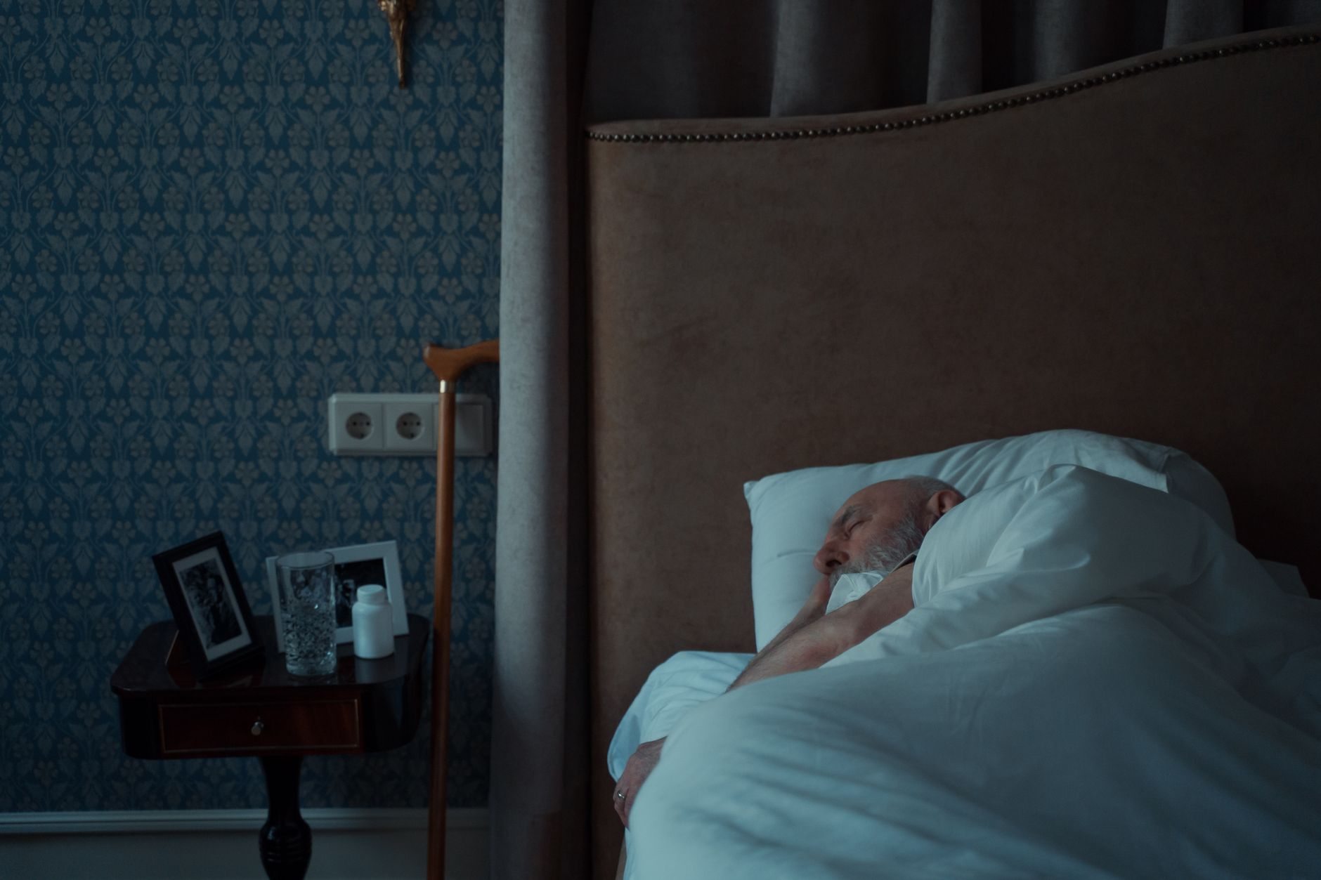
    Yes, Parkinson’s tremor typically stops or significantly reduces during sleep. For most people with Parkinson’s disease, the characteristic resting tremor that occurs during waking hours—especially the pill-rolling tremor in the fingers—becomes largely absent when they fall asleep. A person who experiences visible hand tremors while watching television or sitting at rest will usually find that tremor disappears once they transition into sleep, particularly during deeper sleep stages.

    This happens because the neural circuits responsible for generating tremor in Parkinson’s disease are less active during sleep. The tremor is fundamentally tied to wakefulness and conscious control. When consciousness shifts during sleep, the abnormal electrical rhythms in the basal ganglia that produce tremor are suppressed, giving people with Parkinson’s a period of relative quiet from this symptom. However, the moment a person wakes up, the tremor often returns within seconds or minutes.

    Table of Contents

    Why Does Parkinson’s Tremor Stop When You Sleep?

    The tremor in Parkinson’s disease originates from overactive neurons in the basal ganglia, particularly in circuits involving the substantia nigra, globus pallidus, and thalamus. During wakefulness, especially during relaxation or concentration, these circuits fire in a synchronized, rhythmic pattern at 4-6 cycles per second—the frequency you see as a visible tremor. Sleep disrupts this synchronization because the brain’s electrical patterns change fundamentally. During most sleep stages, the coordinated firing that produces tremor breaks down, and neurons shift into sleep-specific activity patterns.

    Additionally, the neurotransmitter systems that support wakefulness—including dopamine, norepinephrine, and acetylcholine—shift during sleep. Even though dopamine deficiency is central to Parkinson’s disease, the reduction in certain arousal-related systems during sleep removes some of the conditions that amplify tremor. A person with Parkinson’s might notice their tremor is worst when they’re anxious or concentrating, and best during relaxation, and this pattern extends into sleep, where the ultimate relaxation produces near-total tremor suppression. This does not mean sleep “cures” the underlying Parkinson’s pathology, but it temporarily silences one of its most visible symptoms.

    How Tremor Changes Across Different Sleep Stages

    Parkinson’s tremor suppression varies across the sleep cycle in subtle but measurable ways. During light sleep (stages N1 and N2), tremor is usually reduced but may not disappear completely, especially in people with more severe disease. Once a person enters slow-wave sleep (stage N3, also called deep sleep), tremor typically becomes undetectable. However, tremor may briefly re-emerge during REM (rapid eye movement) sleep, when the brain becomes more activated and closer to waking consciousness, though it is usually still reduced compared to wakefulness.

    One important limitation is that not all people with Parkinson’s experience complete tremor suppression during sleep, particularly in advanced stages. Some individuals retain mild tremor even during deep sleep, or experience fragmented sleep patterns that prevent them from reaching deeper stages where suppression is most complete. Sleep quality itself directly affects tremor control—someone with Parkinson’s who has poor sleep architecture (frequent micro-arousals, light fragmented sleep) may experience tremor that returns more readily during the night. This is one reason why sleep disturbances are a significant concern for people with Parkinson’s: poor sleep not only causes daytime fatigue but also prevents the nervous system from fully suppressing tremor and other symptoms.

    Tremor Suppression Across Sleep Stages in Parkinson’s DiseaseLight Sleep (N1-N2)50% reduction from waking baselineDeep Sleep (N3)85% reduction from waking baselineREM Sleep60% reduction from waking baselineWakefulness (Resting)0% reduction from waking baselineWakefulness (Stressed/Anxious)15% reduction from waking baselineSource: Compiled from polysomnography studies and accelerometer tremor tracking in Parkinson’s disease populations

    The Morning Tremor Rebound Effect

    When a person with Parkinson’s wakes up, tremor typically returns quickly—often within seconds to a few minutes. This sudden return is called the morning tremor rebound or sleep-related rebound phenomenon. A person might wake up with hands tremoring at the same intensity as the night before, or sometimes even more pronounced, because the nervous system rapidly re-engages the tremor-generating circuits. This rebound can be particularly noticeable first thing in the morning, before medication has taken effect, making the first 30-60 minutes after waking especially difficult.

    The intensity of the rebound depends partly on medication timing. If someone takes their first dose of levodopa or dopamine agonist right before bed, the medication’s effect might wear off during the latter part of sleep, meaning they wake up in a “medication off” state—which is when tremor is typically worst. Some people find that spacing their evening medication differently, or taking a dose right upon waking, reduces the severity of morning tremor rebound. However, there is a tradeoff: taking medication later in the evening can disrupt sleep itself or cause other nighttime symptoms, so adjustments must be balanced against sleep quality.

    How Parkinson’s Medications Interact With Sleep and Tremor

    The medications used to treat Parkinson’s tremor—levodopa, dopamine agonists like ropinirole or pramipexole, and anticholinergics like trihexyphenidyl—all have effects that extend into sleep and morning periods. Levodopa typically has a half-life of 60-90 minutes, meaning its tremor-suppressing effects wear off relatively quickly during the night. A person taking levodopa at 6 p.m. will likely have minimal medication effect by midnight, and virtually none by 6 a.m. the next morning.

    This is why some people experience significantly worse tremor in the early morning hours and immediately upon waking. Dopamine agonists, which have longer half-lives (often 12-24 hours depending on the specific medication and formulation), provide more sustained coverage through the night and early morning. However, these medications can cause side effects that disrupt sleep, including restless leg syndrome exacerbation, vivid dreams, or delayed sleep onset. There is also a limitation with long-acting formulations: they prevent extreme fluctuations in tremor severity, but some people report they feel less able to notice when medication is working, making it harder to distinguish between their baseline symptoms and medication effects. The goal is usually to find a medication schedule that controls tremor during waking hours while allowing adequate sleep at night.

    When Tremor Doesn’t Stop During Sleep: Complications and Exceptions

    While tremor suppression during sleep is the typical pattern, there are conditions and situations where it doesn’t occur. REM sleep behavior disorder (RBD), which is more common in people with Parkinson’s disease than in the general population, can cause people to physically act out their dreams. During RBD episodes, muscle control returns during REM sleep in an abnormal way, and visible tremor or other involuntary movements can persist or intensify. Additionally, some people with Parkinson’s develop dystonia—sustained muscle contractions—that, unlike tremor, does not reliably stop during sleep and may even worsen at night or early morning.

    Advanced Parkinson’s disease can also change tremor patterns. In later stages, as rigidity and bradykinesia become more prominent, tremor sometimes decreases overall, but sleep-related tremor patterns may become less predictable. Some individuals develop sleep-related hypokinesia, where the inability to move freely during sleep leads to abnormal postures that can trigger pain or other complications. A person with advanced Parkinson’s should mention any changes in nighttime tremor or muscle activity to their neurologist, as these can indicate disease progression or medication adjustment needs. Warning: sudden changes in sleep-related symptoms, such as tremor that no longer stops during sleep, may warrant evaluation to rule out other neurological conditions or medication interactions.

    Sleep Position and Environmental Factors

    The position someone sleeps in can subtly affect tremor suppression, though this is a smaller effect than the fundamental sleep-stage changes. People who sleep on their affected side may experience slightly more tremor awareness when shifting position during sleep (though actual tremor is still suppressed), while sleeping on the unaffected side may feel more comfortable. Some people find that their tremor feels worst when they first lie down and are still semi-conscious, before they fully enter sleep. This is because the transition to sleep is gradual, and tremor suppression increases as sleep deepens.

    Temperature and sleep environment can indirectly affect tremor control by influencing sleep quality. A person with Parkinson’s who sleeps in a room that is too warm may experience fragmented sleep, which prevents deep sleep and therefore prevents full tremor suppression. Conversely, someone who is cold may experience muscle tension that slightly amplifies tremor when they do wake. While these environmental effects are minor compared to the fundamental neurological suppression that occurs during sleep, they are worth considering as part of overall sleep hygiene.

    Tracking Tremor Patterns Across the Sleep-Wake Cycle

    Many people with Parkinson’s and their caregivers find it useful to observe and document their tremor patterns across a 24-hour cycle, including during sleep. Tremor is often less noticeable during the actual sleep period (obviously, since the person is asleep), but it becomes very apparent in the 30-60 minutes after waking. Some people track this by rating their tremor severity at specific times: upon waking, 30 minutes after waking, after taking their first medication dose, and at other consistent times throughout the day.

    This information can help a neurologist adjust medication timing or dosing to better manage morning symptoms. Wearable accelerometers and specialized tremor-tracking devices can measure tremor objectively during sleep and wakefulness, showing exactly how much suppression occurs during different sleep stages. While these are typically used in research settings rather than routine clinical care, they have documented that tremor suppression during sleep is real, quantifiable, and quite profound in most people with Parkinson’s—often a 70-90% reduction from waking levels. Understanding this pattern helps people with Parkinson’s recognize that their nervous system does have periods of relief, and that sleep disruptions can amplify their daytime tremor severity.


    You Might Also Like

  • Can Parkinson’s Tremor Affect the Head?

    Can Parkinson’s Tremor Affect the Head?

    Yes, Parkinson’s tremor commonly affects the head. Head tremor occurs in roughly 5-10% of people with Parkinson’s disease as a primary symptom, though some experience it as the condition progresses. A person might notice their head nodding slightly, especially when they’re concentrating or at rest, or they might feel a subtle shaking side-to-side that worsens with stress or fatigue. This head tremor can be “no-no” (side-to-side shaking) or “yes-yes” (vertical nodding), and it often moves independently from hand or arm tremor.

    Head tremor in Parkinson’s differs from the classic pill-rolling tremor of the hands. It’s driven by the same underlying motor dysfunction—loss of dopamine-producing neurons in the substantia nigra—but manifests differently depending on which neural circuits are most affected. For many people, head tremor is less noticeable than limb tremor and may not cause significant functional problems. For others, especially if combined with neck muscle rigidity, it becomes a visible symptom that draws social attention and affects posture and balance.

    Table of Contents

    What Types of Head Tremor Occur in Parkinson’s?

    parkinson‘s head tremor typically appears as one of two patterns. Anteroposterior tremor (the “yes-yes” motion, nodding forward and backward) is less common but more socially noticeable. Lateral tremor (the “no-no” motion, side-to-side shaking) is the more frequent presentation. Some people experience both, or the tremor pattern shifts over time as the disease progresses. Rest tremor is the most frequent type in Parkinson’s overall, and the head can shake even when completely still.

    This is different from essential tremor, which typically worsens during intentional movement (like holding a cup). With Parkinson’s, you might be sitting quietly and feel your head beginning to shake, then it subsides when you stand and start walking. The frequency is usually slower than hand tremor—typically 4-6 Hz compared to 8-10 Hz in the hands—which can make head tremor feel more pronounced and harder to control consciously. Some people experience task-specific head tremor that only appears during certain activities like writing or eating, similar to how hand tremor can worsen when trying to perform deliberate movements. This mixed presentation makes head tremor unpredictable and harder to manage with a single medication adjustment.

    Visible Impact and Social Recognition

    Head tremor has a distinct disadvantage compared to limb tremor: it’s immediately visible to others. A person shaking their hand can sometimes keep it in their lap or pocket, but head tremor is literally at face level. This visibility often causes more emotional distress than the tremor’s actual functional impact. Studies show that people with noticeable head tremor report higher anxiety in social settings and more frequent comments from strangers about whether they’re cold, nervous, or ill.

    The social impact creates a real feedback loop. Anxiety and stress worsen Parkinson’s tremor, so heightened self-consciousness during social interactions can amplify the head shaking itself. A person might avoid public speaking, dining out, or prolonged eye contact specifically because of visible head tremor, even if their cognitive abilities and speech clarity are unaffected. This can lead to social withdrawal that progresses independently from the underlying motor disability.

    Frequency of Tremor Types in Parkinson’s DiseaseHand/Arm Tremor75%Head Tremor8%Leg Tremor12%Jaw Tremor5%Combined Tremor40%Source: Parkinson’s Disease Foundation; multiple prevalence studies (patients may have overlapping tremor types)

    Head Tremor Versus Limb Tremor: Key Differences

    Head tremor and hand tremor in Parkinson’s share the same neurological origin but behave quite differently. Hand tremor is typically worse at rest and can often be controlled or reduced by voluntary movement—picking up a glass or writing usually suppresses it. Head tremor, by contrast, is less predictably suppressible. You cannot easily “ignore” or “work around” your own head shaking the way you might position a tremoring hand out of sight.

    The frequency and amplitude also diverge. Hand tremor in Parkinson’s is generally faster (8-12 Hz) and occurs in a smaller range of motion, so it might appear as rapid finger twitching. Head tremor is slower (4-7 Hz) and covers a larger arc, making it feel more exaggerated and harder to keep still. This difference affects how medications work: a dose that significantly reduces hand tremor might barely touch head tremor, requiring separate adjustments or additional medication layers. Limb tremor often responds well to dopamine replacement therapy early on, while head tremor sometimes proves more resistant to levodopa alone and may require the addition of other classes of medication.

    Medications and Treatment Approaches

    Levodopa (carbidopa-levodopa) remains the first-line medication for Parkinson’s motor symptoms, including head tremor. However, head tremor responds less consistently than limb tremor. Some people see marked improvement; others notice minimal change even at higher doses. The timing also matters—tremor might decrease during the “on” period when medication is working, but peak during “off” periods when the dose is wearing off or hasn’t yet taken effect. For tremor specifically, anticholinergic medications like benztropine or trihexyphenidyl can be effective adjuncts, particularly if the tremor is prominent relative to other symptoms.

    These drugs are typically used cautiously in older adults due to side effects like confusion and urinary retention, but in younger people with Parkinson’s, they sometimes provide substantial tremor relief. A neurologist might recommend adding an anticholinergic if the head tremor is distressing and levodopa adjustment hasn’t helped. Beta-blockers (like propranolol) and other non-Parkinson’s medications are occasionally prescribed off-label for tremor relief when standard Parkinson’s drugs fall short. Deep brain stimulation (DBS) is an option for advanced disease or tremor-dominant presentations; it can reduce head tremor substantially, though it requires surgery and ongoing device management. The trade-off is significant: DBS offers lasting relief but commits the person to a surgical procedure with potential risks and the need for device adjustments throughout life.

    Progression and Worsening Factors

    Head tremor often becomes more pronounced during early-to-mid disease stages but may stabilize or even improve in later stages if bradykinesia and rigidity become the dominant symptoms. This unpredictability frustrates many people, who assume tremor will worsen steadily and are sometimes relieved (and sometimes alarmed) when the pattern changes. A person with noticeable head tremor in year two of Parkinson’s might find it nearly gone by year five, replaced by stiffness and slowness instead. Stress, fatigue, caffeine, and cold temperatures predictably worsen head tremor acutely. This is critical for caregivers to recognize: the tremor is not a sign the person is nervous or scared—it’s a direct physiological response.

    A person whose head tremor spikes when anxious is not anxious because of the tremor; the tremor worsens because anxiety triggers increased neural firing. This distinction matters for coping: recognizing tremor as a symptom of the disease state, rather than an emotional or behavioral choice, reduces shame and secondary anxiety. Medication “wearing off” (dyskinesias and fluctuations) can also exacerbate head tremor. As Parkinson’s advances, the duration of each levodopa dose shortens, and people experience periods of incomplete symptom control. Head tremor can be one of the earliest signs that the current dose timing or amount needs adjustment.

    Speech and Jaw Effects

    Head tremor often coexists with jaw tremor and speech disturbances in Parkinson’s. When the tremor involves the jaw, it can interfere with speech clarity by introducing involuntary movement into the articulation muscles. A person might sound slightly stuttering or have breaks in their speech flow, not from neurological speech weakness alone but from the jaw shaking beneath the words. Voice quality can also be affected. If the head and neck are shaking, the larynx moves involuntarily, which can make the voice sound tremulous or unstable.

    Some people describe their voice as “shaky” or “wispy” when head tremor is active. This is separate from the hypokinetic dysarthria (weak, quiet, monotone speech) that’s more typical of Parkinson’s overall. The combination can make communication significantly harder: not only is the volume reduced and speed affected, but the perceived tremor in the voice adds another layer of difficulty for listeners to understand. Speech therapy can help, focusing on breath support, articulation exercises, and strategies to stabilize the voice through conscious control. However, there’s a limit: voluntary stabilization gets exhausting and isn’t sustainable for a full day of conversation.

    Functional Impacts on Eating and Vision

    When head tremor is present during eating, the difficulty goes beyond the hand tremor that might make lifting a spoon harder. Head movement can make it harder to guide food into the mouth, and some people find that involuntary head shaking causes them to miss their mouth slightly or drool more, particularly if the tremor is pronounced. This can make mealtimes slower and more effortful, adding another reason to reduce eating in public. Vision can be affected if head tremor is severe enough. The eyes work to stabilize gaze (a reflex called the vestibulo-ocular reflex), but violent or sustained head tremor can overwhelm this system.

    A person might report blurred vision or difficulty reading when their head is shaking, even if the eyes themselves are healthy. The tremor essentially moves the visual field constantly, making it harder to focus on a stable point. For people who also have Parkinson’s-related vision problems (reduced contrast sensitivity, difficulty with bright lights), head tremor compounds these issues. Some people compensate by holding their head more rigidly, using neck muscles to fight the tremor. This leads to secondary problems: neck tension, headaches, and eventually postural strain that affects their overall balance and fall risk.

    Frequently Asked Questions

    Is head tremor a sign my Parkinson’s is getting worse?

    Not necessarily. Head tremor can appear early, remain stable, or even improve as the disease progresses, depending on which brain regions are most affected. Its presence doesn’t predict overall disease severity.

    Why does my head tremor get worse when I’m stressed or tired?

    Stress and fatigue increase neural activity and reduce the brain’s ability to regulate movement. Since Parkinson’s already impairs the motor control circuits, additional stress pushes the system further out of balance, amplifying the tremor temporarily.

    Can head tremor be treated differently than hand tremor?

    Yes. While levodopa helps both, head tremor is often more resistant and may require different medication combinations or higher doses. Some medications effective for hand tremor, like anticholinergics, are specifically used when head tremor is the primary problem.

    Does head tremor affect my thinking or cognitive abilities?

    No. Head tremor is a motor symptom and does not reflect cognitive function. Some people with prominent head tremor have completely normal cognition; others develop cognitive changes separately from the tremor.

    Will deep brain stimulation stop my head tremor?

    DBS can reduce head tremor significantly, especially if tremor is the dominant symptom. However, results vary, and DBS requires surgery and ongoing management. It’s not the first-line option but is considered when medication and other approaches have limited effect.

    Should I avoid social situations because of visible head tremor?

    Head tremor is a recognizable symptom of Parkinson’s and is nothing to be ashamed of. Many people continue social activities and find that their relationships are based on much more than a visible movement disorder. Limiting life due to tremor often increases anxiety and worsens symptoms, creating an unnecessary spiral. —


    You Might Also Like

  • Can Parkinson’s Tremor Affect the Jaw?

    Can Parkinson’s Tremor Affect the Jaw?

    Yes, Parkinson’s tremor can affect the jaw, though this symptom receives far less attention than hand tremor despite affecting roughly 20-30% of people with Parkinson’s disease. Jaw tremor in Parkinson’s—called orofacial tremor—occurs when the same dopamine deficit that causes hand tremor extends to the muscles controlling the jaw and lower face. One patient described the sensation as feeling like his jaw was “chattering involuntarily” during conversations, creating visible movement that made him self-conscious in social settings.

    The jaw tremor in Parkinson’s typically manifests as a side-to-side or vertical up-and-down motion of the lower jaw that becomes more noticeable during stress or fatigue. Unlike essential tremor, which worsens during purposeful movement, Parkinson’s jaw tremor often occurs at rest and can paradoxically worsen when the person tries to consciously control it. The tremor can range from barely perceptible to significantly disruptive, depending on the individual and where they are in disease progression.

    Table of Contents

    How Does Parkinson’s Affect the Jaw and Facial Muscles?

    The mechanism behind jaw tremor involves damage to dopamine-producing neurons in the substantia nigra, a brain region coordinating movement across multiple muscle groups. When dopamine levels decline, the signals regulating smooth jaw movement break down, leaving the muscles of mastication—those responsible for chewing and jaw positioning—susceptible to the same dysregulation affecting the hands, legs, and trunk. This explains why jaw tremor often appears as part of a broader tremor pattern rather than in complete isolation.

    The tremor frequency in jaw muscles typically ranges from 4 to 6 cycles per second, making it visible to others and requiring conscious effort to control. Some people experience asymmetrical jaw tremor where one side shakes more prominently than the other, potentially causing uneven wear on teeth and stress on the temporomandibular joint (TMJ)—the hinge connecting jaw to skull. This asymmetry can create discomfort extending into the neck and ear region.

    Recognizing Jaw and Orofacial Tremor Patterns

    Jaw tremor presents differently across individuals. Some experience rhythmic grinding with audible clicking, while others report a subtle tremor visible mainly when attempting to hold the jaw still. The tremor may involve the lips or tongue, creating a combination of facial movements.

    A significant limitation is that jaw tremor can be mistaken for anxiety-related teeth grinding (bruxism) or TMJ dysfunction, potentially delaying recognition as a Parkinson’s symptom and leading to ineffective treatments targeting the wrong cause. Speech changes frequently accompany jaw tremor, affecting articulation and making speech sound slurred or rushed—a condition called hypokinetic dysarthria. The abnormal jaw movement disrupts the precise tongue-jaw coordination needed for clear speech, particularly in sentences requiring rapid articulation. This differs from Parkinson’s speech difficulties caused by vocal cord stiffness alone, making it important to identify jaw involvement for proper management.

    Orofacial Symptoms in Parkinson’s Disease: PrevalenceJaw Tremor28%Speech Changes60%Drooling50%Difficulty Chewing45%Lip Tightness35%Source: Parkinson’s Foundation Clinical Registry

    The Impact on Eating and Speaking

    Jaw tremor creates genuine practical obstacles during meals. Chewing becomes inefficient when the jaw isn’t stable, as tremor disrupts the rhythmic coordination needed to break down food. Many people with Parkinson’s and jaw tremor report that softer foods become necessary not just due to swallowing difficulty but specifically because the tremor makes chewing harder foods exhausting and time-consuming.

    One man found he needed to choose between his preferred diet and spending twice as long at mealtimes due to tremor-related chewing difficulty. Speaking deteriorates as the day progresses, partly because jaw tremor increases with fatigue and stress. A woman working in client-facing roles noticed that by late afternoon, her jaw tremor became pronounced enough that colleagues remarked on her speech clarity, creating additional social anxiety around a purely physical symptom. This fatigue connection means that timing medication doses around important speaking engagements can be a useful practical strategy.

    Managing Jaw Tremor Through Available Treatments

    Levodopa and dopamine agonists—primary medications for Parkinson’s motor symptoms—often reduce jaw tremor when effective for other tremor types. However, jaw tremor frequently responds less dramatically to medication than hand tremor does, sometimes requiring dose adjustments or timing changes to achieve meaningful relief. This presents a key tradeoff: increasing dopaminergic medication to target jaw tremor might introduce dyskinesias (involuntary writhing movements) elsewhere in the body, forcing patients to balance tremor reduction against new side effects.

    Deep brain stimulation (DBS) shows promise for orofacial tremor, particularly when tremor is the predominant motor symptom. Subthalamic nucleus stimulation can reduce jaw and lip tremor by 50-70% in some patients, though individual response varies considerably. For those not candidates for DBS, adjunctive medications like beta-blockers or anticholinergics may provide modest benefit, though anticholinergics carry particular risks in older patients—including cognitive effects and urinary problems—that must be weighed against tremor relief.

    Complications and When Jaw Tremor Worsens

    Jaw tremor typically worsens during disease progression, becoming one of the more disruptive motor symptoms in middle and later stages. A critical warning: jaw tremor combined with other orofacial motor changes increases aspiration risk—the entry of food or liquid into the airway—because tremor disrupts the coordinated muscle contractions that normally protect the airway during swallowing.

    While not universal, this complication deserves monitoring, especially when jaw tremor coincides with other swallowing difficulties. Chronic jaw clenching from tremor can trigger TMJ disorders, causing pain that radiates to the ear or triggers tension-type headaches. Over time, this muscle tension creates a secondary problem distinct from the Parkinson’s tremor itself, sometimes requiring intervention from a dentist or physical therapist who may initially focus on the pain rather than its neurological source.

    How Stress and Fatigue Amplify Jaw Tremor

    Emotional stress consistently amplifies jaw tremor in Parkinson’s, similar to stress worsening other parkinsonian symptoms. A patient undergoing stressful project deadlines noticed her jaw tremor became nearly continuous during high-pressure weeks, then noticeably decreased during vacation.

    This pattern reflects stress-driven increases in sympathetic nervous system activity, which can magnify tremor independent of disease progression itself. Sleep deprivation particularly magnifies jaw tremor upon waking, with many people reporting most severe tremor in the morning before medication takes effect or after poor sleep. This temporal pattern offers some room for preventive action through prioritizing sleep quality and stress management.

    Dental and Oral Health Concerns

    People with Parkinson’s and jaw tremor need more frequent dental monitoring because tremor causes accelerated, uneven tooth wear on the molars. The grinding pattern from tremor differs from intentional bruxism, sometimes requiring more aggressive dental intervention earlier than expected.

    Dentists unfamiliar with Parkinson’s-related tremor may misdiagnose this wear as intentional grinding and recommend a night guard, which can provide modest help but doesn’t address the neurological cause. Maintaining oral hygiene becomes challenging when jaw tremor affects toothbrush control and coordination. Electric toothbrushes with larger, custom-molded handles reduce self-care frustration, while more frequent professional cleanings manage plaque buildup that tremor-related cleaning difficulties might exacerbate.


    You Might Also Like

  • Can Parkinson’s Tremor Affect the Legs?

    Can Parkinson’s Tremor Affect the Legs?

    Yes, Parkinson’s tremor can affect the legs, though leg involvement is less common than hand tremor. While the characteristic hand shaking associated with Parkinson’s disease gets the most attention, tremors can develop in the feet, ankles, and lower legs in a meaningful percentage of people with the condition. Someone might notice their foot shaking when sitting, or feel vibration in their calf while resting—these can be early signs that Parkinson’s is affecting the lower body. Leg tremor doesn’t always occur in isolation.

    A person might have had hand tremor for years before tremor develops in the legs, or both may appear around the same time. The experience varies significantly from person to person. Some develop tremor in one leg only, while others notice it bilaterally. Understanding how Parkinson’s tremor presents in the legs helps distinguish it from other movement issues and can guide conversations with healthcare providers about symptom management.

    Table of Contents

    Is Leg Tremor Common in Parkinson’s Disease?

    Leg tremor occurs in roughly 25 to 35 percent of people with Parkinson’s disease at some point in their disease course, making it less universal than hand tremor but far from rare. Not everyone with Parkinson’s develops leg tremor—some people experience only upper-body tremor throughout their condition, while others never develop tremor in any limb. The pattern depends on individual disease progression, which can vary dramatically even among people diagnosed around the same age.

    When leg tremor does appear, it often follows the asymmetric pattern typical of early Parkinson’s. A person might notice tremor in the right foot while the left remains unaffected, or vice versa. Over months or years, the tremor may spread to the other leg, remain one-sided, or disappear while hand tremor increases. This unpredictability can make early diagnosis challenging because people sometimes attribute isolated foot tremor to other causes—nerve problems, circulation issues, or muscle fatigue—before recognizing it as part of their Parkinson’s presentation.

    How Does Leg Tremor Feel and Look?

    Parkinson’s leg tremor typically manifests as a visible shaking in the foot or lower leg when the limb is at rest—for example, when sitting on the couch with feet flat on the floor, or lying in bed. The shaking is usually fine to moderate in amplitude, meaning it’s clearly visible but not violent. Someone watching might see the foot bouncing, the calf muscle visibly quivering, or the entire lower leg moving back and forth in a rhythmic pattern. The frequency is typically 3 to 6 cycles per second, similar to hand tremor. One important limitation of leg tremor is that it often diminishes or disappears entirely during intentional movement.

    When someone stands up and walks, the tremor typically stops because walking engages the affected muscles. This is an important diagnostic feature that distinguishes Parkinson’s tremor from other neurological conditions—the tremor emerges at rest and vanishes with purposeful action. However, some people report a peculiar sensation in the resting leg: a buzzing, vibration, or internal shaking that others cannot see. Warning: some people misinterpret this internal sensation as a blood clot or circulatory problem and seek unnecessary vascular workup. It’s worth mentioning this specifically to your doctor.

    Prevalence of Tremor Location in Parkinson’s DiseaseHand Only45%Leg Only12%Hand and Leg22%Tremor-Free21%Source: Parkinson’s Foundation Patient Registry Data (approximated from symptom surveys)

    Leg Tremor Versus Other Movement Changes in Parkinson’s Disease

    Parkinson’s causes several types of movement problems beyond tremor, and leg tremor can coexist with rigidity and bradykinesia in the same limb. Rigidity in the leg feels like stiffness or resistance when someone tries to bend or straighten the knee or hip—imagine moving through honey. Bradykinesia manifests as slowness and reduced amplitude of movement, so a person might take shorter steps or move their leg deliberately and with effort. A person could simultaneously have tremor in the resting foot, rigidity when attempting leg movement, and slowness in walking.

    This combination makes walking more challenging than leg tremor alone would suggest. Comparison: if leg tremor is the visible vibration, rigidity is the resistance you feel, and bradykinesia is the reduced speed and range. A person might describe the experience like this—”My foot shakes when I sit, but when I try to stand or walk, it feels heavy and stiff, and I move slower than I used to.” Tremor often improves preferentially with dopamine medications, while rigidity and bradykinesia also respond but sometimes less dramatically. This difference in medication response can help determine whether tremor or another movement problem is driving a particular functional limitation.

    Managing Tremor in the Legs: What Works and What Doesn’t

    Levodopa and dopamine agonists are the primary medications that reduce Parkinson’s tremor, including leg tremor, in many people. Doses and medication choices that effectively control hand tremor often benefit leg tremor as well, though the leg tremor sometimes responds more slowly or incompletely than hand symptoms. Someone might see their hand tremor disappear entirely on their current medication while mild foot tremor persists, or vice versa. Adjusting the timing of doses—taking medication earlier in the day for tremor that worsens by evening, for example—can help optimize tremor control throughout the day.

    A practical tradeoff: some tremor management strategies that work for the hands are less feasible for the legs. Using weighted utensils or writing aids helps hand tremor by adding inertia, but wearing weighted ankle bands or foot sleeves is cumbersome and provides minimal benefit. Physical therapy focusing on leg strengthening, balance training, and movement patterns can improve function and sometimes reduce the internal sensation of tremor even when the visible shaking persists. One comparison: medication often reduces the amplitude of tremor, while therapy addresses the functional impact—you might still have visible tremor, but you’re more confident walking and less bothered by the sensation.

    When Leg Tremor Gets Worse: Advanced Stages and Complications

    Leg tremor intensity can fluctuate day to day and even hour to hour, influenced by stress, fatigue, caffeine, and disease progression. In some people, leg tremor remains stable for years; in others, it worsens noticeably over months. A warning: some people interpret worsening tremor as a sign that their current medication is failing and request changes urgently. While medication adjustments are sometimes appropriate, worsening tremor can also reflect normal disease progression, changes in sleep quality, or new sources of stress. It warrants evaluation by your neurologist, but it’s not necessarily an emergency.

    Late-stage Parkinson’s sometimes produces tremor that becomes difficult to control with standard medications. In advanced disease, tremor can interfere with nighttime rest or make sitting uncomfortable for long periods. Some people develop secondary complications—muscle fatigue in the leg from constant tremor, or skin irritation if the tremor causes the foot to rub repeatedly against a shoe. These secondary issues sometimes warrant attention even when the tremor itself is minor. Deep brain stimulation, while primarily indicated for tremor that significantly impairs function or for motor complications like dyskinesias, can be considered in specialized cases where leg tremor contributes substantially to disability.

    Medication Effects on Leg Tremor

    Not all anti-Parkinson’s medications affect tremor equally. Levodopa generally provides robust tremor reduction, while dopamine agonists like pramipexole and ropinirole work well for some people but not others. MAO-B inhibitors and COMT inhibitors help manage off-time and motor fluctuations but are not primarily tremor-fighting drugs. Someone whose leg tremor responds well to their current levodopa dose might experience breakthrough tremor during off-periods—times when medication wears off between doses. Adjusting the dosing schedule or adding a longer-acting medication can address this pattern.

    A concrete example: a person taking levodopa three times daily might find that by late afternoon, hand tremor returns mildly but leg tremor returns more noticeably. Their neurologist might prescribe an extended-release formulation or add a dopamine agonist to smooth out medication levels. After this adjustment, both hand and leg tremor remain controlled throughout the day. Not everyone achieves complete tremor elimination—some reach a point where medication reduces tremor by 70 or 80 percent but doesn’t eliminate it entirely. This partial response is common and usually still functional.

    Bilateral Versus Unilateral Leg Tremor in Parkinson’s

    Early Parkinson’s typically features asymmetric symptoms, including one-sided tremor. Someone might have tremor only in the right foot for the first two or three years, with the left leg unaffected. Over time, the tremor often spreads to the other side, though it may remain more prominent on the originally affected side. Bilateral leg tremor—tremor in both feet—occurs in many people with moderate to advanced Parkinson’s, though the severity on each side can differ.

    The asymmetry has practical implications for daily life. A person with right foot tremor might favor their left leg when sitting, crossing their right leg or tucking the right foot under their thigh to reduce visible shaking. As the left leg eventually develops tremor, this compensation strategy becomes less effective. Some people experience tremor that affects one leg primarily during certain times of day or activities, creating an inconsistent pattern that can be confusing. The progression from unilateral to bilateral symptoms typically occurs gradually over months or years, not suddenly, so people usually have time to adjust their strategies and routines.


    You Might Also Like

  • Why Parkinson’s Tremor Often Starts in One Hand

    Why Parkinson’s Tremor Often Starts in One Hand

    Parkinson’s tremor typically starts in one hand because dopamine-producing neurons in the substantia nigra—a region deep in the brain—degenerate unevenly. Early in the disease, cell death concentrates in one area of this structure, affecting the brain circuits that control movement on the opposite side of the body. If a person develops tremor in their right hand, it usually signals that the left side of their substantia nigra has begun losing dopamine-producing cells. This asymmetrical pattern is one of the disease’s defining early characteristics and can often be traced back through years of subtle motor changes that precede the obvious shaking.

    The hand where tremor appears is not random. In about 75% of Parkinson’s cases, the tremor remains confined to one limb for months or even years before potentially spreading to the opposite side. A person might notice their right hand shaking during rest while the left hand moves normally—sometimes for an extended period before any other symptoms emerge. This unilateral onset is so consistent that doctors often look for it as a clue when evaluating potential Parkinson’s diagnosis.

    Table of Contents

    Why Does Brain Degeneration Start on One Side?

    The human brain’s motor system is lateralized, meaning each hemisphere controls movement on the opposite side of the body. When Parkinson’s disease begins its damage, it does not affect both hemispheres equally. Researchers still do not fully understand why the degeneration concentrates on one side initially—it may relate to differences in how cells were distributed during development, variations in blood flow, or subtle differences in individual brain chemistry. What is clear is that in early Parkinson’s, the substantia nigra on one side loses more dopamine-producing neurons than the other.

    This asymmetry can persist for years. Some people with early-stage Parkinson’s experience tremor only in the right hand or only in the left for 3, 5, or even 10 years before symptoms noticeably emerge on the other side. Unlike conditions where both sides of the brain are equally affected from the start, Parkinson’s typically shows a clear dominance pattern. A person might develop excellent coping strategies for their affected hand—learning to write with their opposite hand or using adapted utensils—only to face a new phase of adjustment when the other side eventually becomes symptomatic.

    The Substantia Nigra’s Role in Movement Control

    The substantia nigra, located in the midbrain, contains roughly 500,000 dopamine-producing neurons in each person. These neurons release dopamine throughout the motor system, enabling smooth, coordinated movement and the ability to initiate motion without tremor. In Parkinson’s disease, these neurons die at an accelerated rate. By the time a person notices tremor in one hand, they have typically lost between 50% and 70% of dopamine-producing cells on that side of the brain—a substantial loss that has unfolded over years, often silently.

    One limitation in early diagnosis is that the correlation between cell loss and visible symptoms is not precise. Two people with similar amounts of dopamine neuron loss might experience very different tremor severity—one might have a barely noticeable vibration while another has obvious shaking. Some people lose enough dopamine cells to cause tremor in one hand while the other hand remains completely unaffected, even though that side of the brain is also experiencing cell death. This mismatch between brain pathology and symptom visibility can cause frustration when neurologists run standard motor tests and find that both hands appear to function normally, even though a person experiences clear tremor when at rest.

    Percentage of Parkinson’s Patients With Unilateral vs. Bilateral Tremor at DiagnUnilateral (One Hand)62%Unilateral (Other Sites)8%Already Bilateral18%No Tremor Symptom12%Source: Parkinson’s Foundation patient registry data; typical presentation patterns in early-stage disease

    Dopamine Loss and the Resting Tremor Pattern

    parkinson‘s tremor characteristically appears when the hands are at rest—not during movement or when gripping an object. This distinctive feature is tied directly to dopamine loss in the substantia nigra. When dopamine levels drop asymmetrically, the brain’s ability to suppress unwanted movements becomes imbalanced. The side with greater cell loss loses this suppression first, resulting in the visible shaking.

    A person might hold their right hand steady while typing but notice noticeable tremor when their hand rests in their lap—this on-off pattern reflects the brain’s shifting demand for dopamine during different motor states. Why does the tremor not always spread to the opposite hand? The answer involves both neurobiology and individual variation. In some people, the degeneration in the substantia nigra remains genuinely asymmetrical for years—one side continues losing cells much faster than the other. In others, both sides are degenerating at similar rates, but the threshold for visible symptoms differs between hemispheres due to differences in neural connectivity or receptor sensitivity. A 60-year-old diagnosed with unilateral Parkinson’s tremor might experience spread to both hands within a year, or might remain predominantly unilateral for 15 years with only subtle changes on the opposite side.

    Detecting and Confirming Unilateral Tremor

    A neurologist typically assesses tremor by watching the hands at rest and during movement, noticing frequency, amplitude, and which side of the body is involved. Parkinson’s tremor has a characteristic frequency of 4 to 6 hertz—slower and more regular than the fine tremor seen in essential tremor or anxiety-related shaking. When tremor appears in only one hand or arm during these observations, it strongly supports a Parkinson’s diagnosis, though other tests help rule out mimics. Comparing Parkinson’s tremor to other tremor types reveals important distinctions.

    Essential tremor, a much more common condition, typically affects both hands symmetrically and worsens with intentional movement or holding a position—exactly opposite to Parkinson’s pattern. A person with essential tremor might struggle to hold a coffee cup steady, while a person with early Parkinson’s might have steady hands while actively holding the cup but obvious shaking when their hands rest on a table. Dystonic tremor, another mimic, often involves abnormal postures or twisting movements alongside the shaking. These differences matter because they guide treatment—medication that helps Parkinson’s tremor may not help essential tremor, and misidentification leads to ineffective or inappropriate therapy.

    Progression From One Hand to Both Hands

    For the majority of people with Parkinson’s disease, the tremor does eventually spread to involve both hands and sometimes other body parts like the jaw, lips, or legs. This spread typically occurs within 2-5 years of initial symptoms, though the timeline varies considerably. A person whose tremor started in the right hand at age 60 might have bilateral tremor by 62 or 64, or might still experience predominantly right-sided tremor at 70 despite having other Parkinson’s symptoms like bradykinesia (slow movement) affecting both sides.

    One important warning: the absence of tremor spreading does not mean the disease is not progressing. Someone with unilateral tremor who remains unilateral for many years can still experience significant worsening of balance, gait, cognitive function, or motor control on both sides of the body. Tremor is only one symptom among many in Parkinson’s disease, and it does not progress in lockstep with other aspects of the condition. A person might have the relief of tremor remaining limited to one hand while contending with increasingly severe stiffness, slowness, or balance problems affecting their whole body.

    Imaging and Structural Evidence

    Modern neuroimaging can reveal asymmetrical dopamine loss even before obvious tremor appears. PET (positron emission tomography) scans using tracers that bind to dopamine can show which side of the substantia nigra has retained more dopamine-producing function. DaT scans (dopamine transporter imaging) show asymmetrical patterns in early Parkinson’s that correlate with which hand has tremor.

    A scan might show that the left substantia nigra has retained 45% of normal dopamine function while the right has retained 70%—a difference that explains why tremor appears in the right hand, which is controlled by the left brain. These imaging studies confirm that unilateral tremor reflects genuine asymmetrical brain pathology, not coincidence or preference. They also show that some people with future Parkinson’s diagnosis have detectable dopamine loss on both sides of the brain before tremor becomes obvious anywhere, meaning the asymmetry visible in symptoms does not always match the exact symmetry of cell loss. Early detection through imaging remains a research tool rather than routine clinical practice, but it underscores that the tremor pattern people notice is downstream from real, measurable changes in the brain.

    Medication Response and Symptom Management

    When tremor remains limited to one hand, dopamine-replacement medications like levodopa typically reduce or eliminate it. A person taking carbidopa-levodopa might notice their tremor diminishes within 30 minutes of taking a dose, and their tremor-free hand allows them to perform tasks like writing or eating more confidently. However, some people experience incomplete tremor suppression—the medication improves the tremor but does not completely stop it, or it stops the tremor but creates new movement side effects.

    Over time, the same medication doses that initially controlled unilateral tremor may become less effective, or the tremor may gradually spread to the other hand despite stable medication doses. Some people adapt by adjusting timing of doses around daily activities, taking medication before activities that trigger tremor awareness. Others manage by learning to position affected limbs in less visible ways or by understanding their tremor rhythms well enough to anticipate when it will be worse—such as when tired, stressed, or during cold weather. The fundamental reality is that unilateral tremor, though limited to one side, still connects to a progressive brain condition that typically involves both sides eventually.


    You Might Also Like

  • What Does a Parkinson’s Resting Tremor Look Like?

    What Does a Parkinson’s Resting Tremor Look Like?

    A Parkinson’s resting tremor characteristically appears as a rhythmic, involuntary shaking of the hands or other body parts when they are at rest. The most distinctive form is the “pill-rolling” tremor, where the thumb and index finger move repetitively in a circular motion as if rolling a small pill or stone between them. This tremor occurs at a frequency of 4 to 6 cycles per second, and it disappears or substantially decreases when the affected person initiates voluntary movement or falls asleep—a feature that makes it diagnostically unique and helps physicians distinguish it from other movement disorders.

    The tremor typically begins on one side of the body, often affecting one arm or leg initially, though it may progress to both sides over time. About 75 percent of people with Parkinson’s disease experience resting tremor at some point during their illness, making it one of the most recognizable symptoms and frequently the first sign that prompts someone to seek medical evaluation. The tremor can vary dramatically in intensity, sometimes barely noticeable and other times severe enough to make fine motor tasks like writing or eating with utensils difficult.

    Table of Contents

    HOW THE PILL-ROLLING TREMOR APPEARS

    The pill-rolling tremor gets its name from the visual appearance of the movement pattern. The thumb makes repeated contact with the index finger in a slow, rhythmic circular motion, creating the optical illusion of rolling something small between the digits. This pattern is so characteristic of Parkinson’s disease that neurologists consider it a hallmark diagnostic sign. The tremor affects the distal portions of the limbs—the fingers, hands, forearms, and lower legs—with greater intensity in the fingers and hands, while more proximal areas like the upper arms and shoulders may be spared entirely. The amplitude, or width of the tremor movement, varies considerably from person to person and even changes throughout a single day.

    Some individuals may have barely perceptible tremors less than 1 centimeter wide, while others experience tremors greater than 10 centimeters in amplitude. One important distinction from other tremor types is that Parkinsonian tremor is entirely absent during sleep and vanishes or nearly vanishes the moment the person engages in purposeful activity. For example, a person with resting tremor in their hands while sitting might see the tremor completely disappear when they pick up a pen to write or reach to open a door. The tremor may also spread to the jaw, tongue, chin, or rarely the trunk, though tremor affecting the head is notably absent in typical Parkinson’s disease and should prompt physicians to reconsider the diagnosis. Unlike essential tremor, which worsens when a person holds their hands in a certain position or during movement, Parkinsonian tremor specifically worsens during periods of mental stress, anxiety, or emotional arousal, and is triggered by inactivity.

    FREQUENCY AND AMPLITUDE PATTERNS

    parkinson‘s resting tremor vibrates at a relatively slow, consistent frequency compared to other tremor disorders. The standard frequency range is 4 to 6 cycles per second (Hz), though medical literature sometimes cites the wider range of 3 to 7 Hz. This frequency is markedly slower than essential tremor, which typically occurs at 8 to 12 Hz and creates a noticeably faster, finer quivering. The slowness of Parkinsonian tremor is one of the clinical clues that helps experienced neurologists identify it at the bedside, and it is often easily visible to observers even from a distance.

    The amplitude of the tremor—how far the affected body part moves with each oscillation—is highly variable and unpredictable. An individual might experience dramatic changes in tremor intensity from hour to hour or even from moment to moment depending on psychological state, fatigue level, and medication timing. Mental concentration or emotional distress amplifies the tremor noticeably, which can be challenging for individuals who must perform fine tasks like signing documents or eating in public. Conversely, engaging in purposeful voluntary movement—whether reaching for an object, writing, or even having a conversation while gesturing—causes the tremor to diminish or vanish entirely.

    Prevalence of Rest Tremor and Other Tremor Types in Parkinson’s DiseaseRest Tremor Only14.5%Rest + Postural18%Rest + Kinetic22%Rest + Both Action Types22%No Tremor25%Source: NIH/PMC peer-reviewed studies on Parkinson’s tremor prevalence; analysis across multiple patient cohorts

    ASYMMETRIC ONSET AND PROGRESSION PATTERNS

    One of the most clinically useful characteristics of Parkinson’s resting tremor is its asymmetric beginning. The tremor almost always starts on one side of the body—typically one arm, though occasionally one leg—rather than affecting both limbs simultaneously. This asymmetric pattern is a diagnostic clue that helps distinguish Parkinson’s disease from other conditions.

    Over months or years, the tremor may spread to the opposite side of the body, but many patients continue to experience more pronounced tremor on the initially affected side even after the disease becomes bilateral. The initial presentation of unilateral tremor raises important practical considerations for diagnosis and management. When a patient first notices unilateral hand tremor, the progression is rarely immediately bilateral; instead, it often remains prominent on one side while the opposite limb remains either unaffected or develops only mild tremor. This asymmetry is so characteristic that when a neurologist observes perfectly symmetrical tremor from the disease’s onset, it may indicate a condition other than idiopathic Parkinson’s disease, such as essential tremor or drug-induced parkinsonism.

    DIAGNOSTIC SIGNIFICANCE AND CLINICAL ASSESSMENT

    Parkinson’s resting tremor holds tremendous diagnostic weight because it serves as a required component in the definition of parkinsonian syndrome and is one of the cardinal features that doctors look for when evaluating a patient suspected of having Parkinson’s disease. The tremor is assessed and documented by neurologists based on several observable criteria: the frequency of oscillation, the amplitude of movement, the specific body parts affected, the degree to which voluntary movement suppresses the tremor, and the response to emotional stress. Physicians performing a neurological examination typically ask patients to relax their hands in their lap or resting on a table, and then they observe the tremor closely and may count its frequency.

    They will then ask patients to reach toward the examiner’s finger or perform other intentional movements to confirm that the tremor resolves with voluntary activity. Unlike action tremor, which worsens during purposeful movement and does not respond as well to dopamine replacement therapy, resting tremor is characteristically suppressed by movement and responds dramatically to L-Dopa medication—a distinction that physicians use both for diagnosis and for predicting treatment response. Pill-rolling tremor appears in only a limited set of conditions, primarily Parkinson’s disease but also potentially in drug-induced parkinsonism (caused by antipsychotic medications or antiemetics that block dopamine), and in rare atypical parkinsonian disorders like progressive supranuclear palsy or multiple system atrophy with parkinsonian features. This specificity makes the observation of classic pill-rolling tremor highly valuable to the diagnostic process.

    PREVALENCE AND LIFETIME EXPERIENCE

    Research data shows that resting tremor occurs in approximately 58 percent of people with Parkinson’s disease based on averaged prevalence figures across multiple patient cohorts. When examining broader tremor experience—including resting, postural, and kinetic tremors of all types—the percentage rises substantially, with 70 to 90 percent of Parkinson’s patients experiencing some form of tremor during their lifetime. Over an extended 7-year follow-up period, 87.2 percent of patients demonstrated rest tremor in at least one clinical assessment, indicating that even patients who may not exhibit tremor at initial diagnosis often develop it eventually.

    Importantly, not all tremors in Parkinson’s patients are pure resting tremors. Only 14.5 percent of patients experience “pure” resting tremor without any postural or kinetic (action) tremor component; most patients have a combination of tremor types. This mixed presentation affects management decisions because different tremor types respond differently to medications and deep brain stimulation surgery. Resting tremor responds more favorably to L-Dopa therapy and to subthalamic nucleus deep brain stimulation (STN-DBS), whereas action tremor tends to be more refractory to dopaminergic treatment and may even worsen progressively despite good control of resting tremor.

    RELATIONSHIP TO DISEASE PROGRESSION AND SYMPTOM EVOLUTION

    While resting tremor is often the earliest movement symptom of Parkinson’s disease and frequently prompts initial medical evaluation, an interesting clinical paradox occurs: tremor prominence may actually decrease as the overall disease progresses. This means that while the person’s Parkinson’s disease may be advancing with worsening rigidity, slowness of movement, and balance problems, the tremor itself might become less visually apparent. This phenomenon is not well understood but appears to relate to broader changes in motor control and basal ganglia function as the disease evolves.

    The initial presentation of tremor often raises the question of prognosis. Patients who first manifest Parkinson’s disease as a tremor-dominant presentation (versus a rigidity-dominant or bradykinesia-dominant presentation) tend to have somewhat different disease trajectories and may experience a slower rate of motor decline than patients with akinetic-rigid presentations. However, tremor-dominant presentations are not predictive of whether the overall disease course will be mild or severe.

    DISTINGUISHING FEATURES COMPARED TO OTHER TREMOR DISORDERS

    Parkinsonian resting tremor differs fundamentally from essential tremor, the most common pathological tremor type in the general population. Essential tremor worsens when the hands are held in a particular position (postural tremor) or during purposeful movement (kinetic tremor), whereas Parkinsonian tremor improves with purposeful movement. Essential tremor is also faster—typically 8 to 12 Hz—and often has a family history, affects both sides of the body relatively symmetrically from onset, and does not respond to dopaminergic medications.

    The distinction between these two conditions is critical because the treatments are entirely different: essential tremor may respond to propranolol or primidone, while Parkinsonian tremor responds to dopamine replacement. Orthostatic tremor, another distinct condition, occurs in the legs and trunk when standing and subsides when sitting or walking—a pattern opposite to Parkinsonian tremor. Cerebellar tremor, arising from damage to the cerebellum, worsens during purposeful movement and is noticeably absent at rest, creating another clear contrast with the rest tremor of Parkinson’s disease.


    You Might Also Like

  • When to See a Neurologist About Possible Parkinson’s Signs

    When to See a Neurologist About Possible Parkinson’s Signs

    You should see a neurologist about possible Parkinson’s signs when you notice motor symptoms that persist for more than a few weeks—tremor at rest, stiffness, slowness of movement, or balance problems—especially if they’re affecting your daily function or appearing on only one side of your body. These aren’t symptoms to investigate alone with your primary care doctor; a neurologist, particularly one with movement disorder expertise, has the training to recognize Parkinson’s-specific patterns that internists often miss. For example, a 58-year-old man noticed his right hand shook when resting on his lap, his handwriting became smaller, and he felt stiff in his right shoulder.

    His family doctor initially attributed it to stress, but when the tremor worsened and he began shuffling when walking, a neurology referral revealed early Parkinson’s disease—caught early enough that lifestyle interventions and medication options could be discussed before symptoms progressed further. The urgency depends on several factors: whether your symptoms are spreading to both sides of your body, whether you’re experiencing cognitive changes alongside motor symptoms, and whether you have a family history of Parkinson’s. A neurologist can order the specific tests and imaging that actually distinguish Parkinson’s from other conditions that mimic it, like essential tremor, drug-induced parkinsonism, or progressive supranuclear palsy. Waiting months or years for a diagnosis while symptoms worsen doesn’t give you time to plan treatment or lifestyle changes—it only allows the disease to progress unchecked.

    Table of Contents

    What Motor Symptoms Signal It’s Time to See a Neurologist

    The classic signs of Parkinson’s disease include a resting tremor (shaking when your hand is relaxed, not moving), rigidity (muscle stiffness that persists throughout movement), bradykinesia (slowness of voluntary movement), and postural instability (difficulty with balance and posture). If you have any combination of these lasting several weeks, especially if they started on one side of your body and haven’t improved, a neurologist appointment should be scheduled soon. Many people dismiss these symptoms as normal aging or stress-related, but Parkinson’s symptoms follow a specific pattern: they typically start asymmetrically (one side first) and progress gradually over months. Non-motor symptoms also warrant neurological evaluation. Constipation, sleep disturbances, loss of smell, and mood changes often precede motor symptoms by years in Parkinson’s disease.

    If you’ve had a recent significant change in your sense of smell, new-onset depression, or sudden constipation alongside any subtle motor changes, these clusters suggest a neurologist should evaluate you. A 62-year-old woman lost her ability to smell coffee and developed depression, which her doctor treated with antidepressants, but when she started experiencing arm stiffness six months later, a neurologist recognized the pattern and diagnosed early-stage Parkinson’s—the smell loss and mood changes were early warning signs all along. The timeline matters significantly. Essential tremor, which is often confused with Parkinson’s tremor, remains stable or worsens very slowly over decades. Parkinson’s symptoms, by contrast, show measurable progression—tremor becomes more pronounced, stiffness spreads, or balance worsens—within weeks to months. If your symptoms are clearly progressing, don’t delay the neurologist visit hoping they’ll improve on their own.

    Risk Factors and When You Shouldn’t Wait

    Age is a major factor: Parkinson’s disease typically starts after age 60, though it can appear earlier. If you’re in your 50s or 60s and developing unexplained tremor or stiffness, earlier evaluation is justified because earlier diagnosis allows for earlier intervention. Family history increases risk significantly—if a parent, sibling, or child has Parkinson’s, your threshold for seeking neurological evaluation should be lower. Environmental exposure to pesticides, herbicides, or living near industrial areas also raises risk, and if you have that exposure history plus motor symptoms, you shouldn’t wait. One critical limitation: some neurologists practice in areas without movement disorder specialists, so availability matters.

    If your local neurologist has a three-month wait and you’re experiencing rapidly worsening symptoms, consider traveling to a tertiary care center or university hospital with a movement disorders clinic. A 54-year-old man with a two-month history of worsening tremor and stiffness lived in a rural area where his neurologist was booked out four months. His symptoms progressed rapidly, making his work and driving increasingly difficult. Had he contacted the nearest university hospital two hours away, he could have been evaluated and started on medication two months earlier, potentially preventing some functional decline. The distinction between “concerning symptoms” and “urgent symptoms” is important. Symptoms that are worsening rapidly (noticeable progression week-to-week rather than month-to-month), causing significant functional impairment, or spreading to both sides of your body quickly deserve urgent attention—call for an expedited appointment rather than accepting a routine slot months away.

    Percentage of Parkinson’s Patients by Age at DiagnosisAge 40-505%Age 51-6015%Age 61-7035%Age 71-8030%Age 81+15%Source: Parkinson’s Foundation Statistics

    The Neurological Examination and Diagnosis Process

    When you see a neurologist for suspected Parkinson’s, they won’t diagnose you based on a single test. Instead, they perform a detailed motor exam looking for the specific combination of signs: resting tremor, rigidity tested through passive movement, bradykinesia observed during timed finger-tapping or rapid hand movements, and postural stability tested with the pull test. They also examine eye movements, facial expression, and gait—Parkinson’s produces a characteristic “masked” face and shuffling gait that experienced neurologists recognize. The exam itself takes 20–40 minutes and provides more diagnostic information than any blood test or brain scan. Imaging tests like MRI or PET scans can rule out other conditions (like structural brain abnormalities or atypical parkinsonism), but they cannot definitively diagnose Parkinson’s disease. That diagnosis is still clinical, based on the neurologist’s assessment of your symptoms and exam findings.

    A neurologist might order dopamine transporter imaging (DaTscan) if the diagnosis is uncertain, but this test is expensive, not always covered by insurance, and isn’t required for most straightforward cases. The neurologist might also refer you for neuropsychological testing if cognitive symptoms are present, since Parkinson’s dementia develops in some patients years after motor onset. A limitation to understand: some neurologists, particularly those without movement disorder subspecialty training, misdiagnose Parkinson’s as essential tremor or vice versa. Essential tremor appears with action (when you’re using your hands), while Parkinson’s tremor appears at rest. If a neurologist doesn’t thoroughly test for this distinction, diagnostic error is possible. Getting a second opinion from a movement disorders specialist is entirely reasonable if your initial diagnosis seems uncertain or if your symptoms don’t fit the expected Parkinson’s pattern.

    Preparing for Your Neurologist Appointment: What to Bring and Know

    Before your appointment, write down your symptoms in order of appearance—when did the tremor start, when did you first notice stiffness, when did your handwriting change? Include how your symptoms have progressed: are they stable, slowly worsening, or rapidly worsening? Note any family history of neurological disease, medications you’re taking (some medications can cause parkinsonism), and any recent exposures to pesticides or industrial chemicals. Bring this information in writing because neurologists review it closely, and details matter for diagnosis. Document your functional changes: Can you still button shirts? Is your handwriting becoming illegible? Are you falling? Is your sleep disrupted? Are you experiencing depression or anxiety? These specific examples help the neurologist understand the disease’s impact. Many patients downplay their symptoms in the moment, but written documentation prevents this.

    A 67-year-old woman brought a video of her tremor, examples of her recent handwriting compared to old samples, and a list of activities she could no longer do—this concrete evidence helped the neurologist immediately recognize Parkinson’s disease where a verbal description alone might have created ambiguity. Bring your complete medication list, including over-the-counter medications and supplements, because some can mimic or worsen Parkinson’s symptoms. Antipsychotics, certain antiemetics (like metoclopramide), and some antidepressants can cause drug-induced parkinsonism, which mimics Parkinson’s disease but may resolve if the medication is stopped. Your neurologist needs to rule this out before making a diagnosis.

    Red Flags and When Neurological Evaluation Becomes Urgent

    Certain presentations require urgent neurological evaluation rather than waiting for a routine appointment. Rapid worsening of motor symptoms over days to weeks, sudden onset of cognitive changes (confusion, memory loss, or personality changes) alongside motor symptoms, or development of severe balance problems that cause falls should prompt a call to your neurologist asking for an urgent appointment or even emergency department evaluation. Atypical presentations like sudden-onset tremor in both hands simultaneously, or tremor that worsens with intentional movement (action tremor) rather than at rest, might indicate a different neurological condition entirely and need rapid evaluation. Falls are a warning sign. If balance impairment develops rapidly or you’re falling frequently, this suggests either advanced Parkinson’s disease or a different condition (atypical parkinsonism, stroke, or normal pressure hydrocephalus).

    A 71-year-old man with early-stage Parkinson’s had been managed for two years without significant balance problems. When he suddenly began falling multiple times weekly, his neurologist discovered he had developed normal pressure hydrocephalus—a separate condition requiring different treatment. Rapid worsening of balance can indicate disease progression, but it can also signal a treatable separate problem, so it demands urgent evaluation. Cognitive decline combined with parkinsonism is also a warning pattern. Parkinson’s disease can eventually include cognitive impairment, but early dementia alongside motor symptoms might indicate Lewy body dementia or another atypical condition. If you’re experiencing confusion, hallucinations, or significant memory loss that developed suddenly or rapidly, this distinction matters because treatment approaches differ.

    Finding a Neurologist with Movement Disorder Expertise

    Not all neurologists specialize in movement disorders, and this specialization matters for accurate diagnosis. A general neurologist can recognize straightforward Parkinson’s disease, but if your presentation is atypical, if you’re younger than typical Parkinson’s patients, or if your diagnosis remains uncertain, you want a movement disorder specialist. These physicians have additional fellowship training specifically in conditions like Parkinson’s, dystonia, and tremor. They’re more likely to catch diagnostic errors and have greater expertise with medication adjustments and advanced therapies. Finding a movement disorders specialist requires some legwork.

    If you live in or near a major city with a university medical center, that center almost certainly has a movement disorder clinic. If you live rurally, you might need to travel, but the investment in getting to a specialist for initial evaluation and diagnosis is worthwhile. Your primary care doctor should be able to refer you, or you can contact your state neurological society for a specialist list. The International Parkinson and Movement Disorder Society maintains a physician directory. If you’re struggling to find a specialist and your diagnosis is urgent, consider asking for a telemedicine consultation with a specialist at a distant institution—many now offer virtual visits.

    Starting Treatment and Ongoing Neurologist Care

    Once diagnosed, your neurologist becomes your ongoing partner in managing Parkinson’s disease. Initial management typically involves discussion of medication options—levodopa/carbidopa is the gold standard, though dopamine agonists and other medication classes are alternatives depending on your age, symptoms, and other health conditions. Your neurologist discusses these options, helps you understand benefits and side effects, and tailors the choice to your individual situation. This conversation requires time and shouldn’t be rushed; a good neurologist spends 30+ minutes on your first treatment discussion. Ongoing neurologist care involves regular follow-up appointments (usually every 3–6 months initially) to assess how medications are working, whether side effects have developed, and whether your symptoms are controlled adequately. As Parkinson’s progresses, medication adjustments become necessary—doses increase, or additional medications are added.

    Your neurologist monitors for complications like dyskinesia (involuntary movements caused by long-term levodopa) or motor fluctuations (periods of better or worse function). They also screen for non-motor complications: cognitive decline, depression, blood pressure changes, and sleep problems. A 72-year-old man was on stable levodopa therapy for three years when his neurologist noticed during routine screening that his blood pressure had become dangerously low—a known complication of Parkinson’s—and adjusted his medications accordingly, preventing falls and hospitalization. Regular, attentive neurologist care catches these complications early. Advanced treatments like deep brain stimulation (DBS) for motor symptoms or new medications for non-motor symptoms become options as disease progresses, but your neurologist must monitor your disease trajectory to recommend these therapies at the right time. This requires continuity of care with a single neurologist or clinic so that disease progression is tracked accurately over years. Changing neurologists frequently or relying on intermittent urgent care visits prevents the longitudinal relationship necessary for optimal management.

    Frequently Asked Questions

    If my primary care doctor thinks my tremor is just stress, should I push for a neurologist referral anyway?

    Yes. Stress doesn’t typically cause resting tremor or progressive stiffness. If your doctor dismisses your symptoms but you’re concerned, request the referral directly. You can also see a neurologist without a referral in many insurance plans—call and ask.

    How quickly should I expect to see a neurologist if I call with possible Parkinson’s symptoms?

    Routine appointments may have a 4–12 week wait, but if you explain that you’re experiencing progressive motor symptoms, many practices can fit you in sooner—within 2–4 weeks. If symptoms are rapidly worsening, ask specifically for an expedited or urgent slot.

    Could my symptoms be something other than Parkinson’s, and how would a neurologist tell the difference?

    Yes—essential tremor, drug-induced parkinsonism, progressive supranuclear palsy, and normal pressure hydrocephalus can all mimic Parkinson’s. A neurologist distinguishes these through careful history, physical exam, and sometimes imaging. This is exactly why seeing a neurologist matters rather than assuming a diagnosis.

    Do I need a brain MRI or scan to diagnose Parkinson’s disease?

    Not necessarily. Parkinson’s is diagnosed clinically through neurological examination. Imaging is used to rule out other conditions (like stroke or brain tumors), but a normal MRI doesn’t exclude Parkinson’s, and an abnormal finding doesn’t diagnose it.

    What should I do if I can’t get a neurologist appointment for several months but my symptoms are worsening?

    Call the office and specifically request urgent or expedited scheduling, explaining that your symptoms are progressing. If that doesn’t work, ask for a referral to another neurologist with sooner availability, or investigate telemedicine consultations with movement disorder specialists at university hospitals.

    Can Parkinson’s be diagnosed before symptoms appear, and should I see a neurologist if I just have family history?

    Currently, there’s no test to diagnose preclinical Parkinson’s. If you have family history but no symptoms, routine neurologist visits aren’t indicated, though discussing your risk with your primary care doctor and staying alert to symptom development is reasonable. If symptoms do appear, then neurologist evaluation is warranted. —


    You Might Also Like

  • Can Sleep Changes Precede Parkinson’s by Years?

    Can Sleep Changes Precede Parkinson’s by Years?

    Yes, sleep changes can precede Parkinson’s disease by years—sometimes a decade or more before motor symptoms like tremor or stiffness appear. Research into rapid eye movement (REM) sleep behavior disorder (RBD) shows that people who act out their dreams during sleep face a significantly elevated risk of developing Parkinson’s disease later. Studies have found that patients with RBD have roughly a 30 to 50 percent chance of being diagnosed with Parkinson’s or another synucleinopathy (a group of neurodegenerative diseases involving abnormal alpha-synuclein protein accumulation) within 10 to 15 years. One patient, James, began thrashing and punching in his sleep at age 48, often waking his wife with sudden movements.

    His sleep specialist noted the RBD diagnosis, and twelve years later, at age 60, he received a Parkinson’s diagnosis—his sleep disorder had been the earliest warning sign that his brain was beginning to degenerate. Sleep disturbances in early Parkinson’s are not limited to RBD. Many people also experience restless leg syndrome, insomnia, excessive daytime sleepiness, and sleep apnea years before they notice a tremor or feel their movements slowing down. The brain changes underlying Parkinson’s disease—specifically the loss of dopamine-producing neurons in a region called the substantia nigra—can disrupt the neural circuits controlling sleep long before they affect the motor system enough to cause noticeable symptoms. This means that sleep disturbances can serve as an early-warning system, offering a window of time for closer monitoring and potentially earlier intervention.

    Table of Contents

    What Sleep Changes Tell Us About Parkinson’s Risk

    Sleep disorders linked to Parkinson’s are not merely a consequence of the disease—they often appear in the prodromal phase, the period before diagnosis when pathology is present but motor symptoms have not yet emerged. RBD is the most studied of these precursor signs. In RBD, the muscle atonia (paralysis) that normally prevents physical movement during REM sleep fails. Instead of being temporarily paralyzed while dreaming, people with RBD act out their dreams, sometimes violently. They might punch, kick, or fall out of bed while experiencing vivid, often unpleasant dreams. Video polysomnography (sleep monitoring with cameras) can confirm the diagnosis by capturing the absence of normal muscle atonia during REM periods combined with abnormal behavior.

    The specificity of RBD for Parkinson’s and related diseases is striking. Unlike many risk factors that predict multiple conditions, RBD carries a particularly high probability of progression to a synucleinopathy. Longitudinal studies following patients with RBD over 10 to 15 years have found that roughly 30 to 50 percent develop Parkinson’s disease, multiple system atrophy, or Lewy body dementia. This makes RBD one of the most reliable early biomarkers we currently have. A woman in her early 50s who was diagnosed with RBD and had repeat sleep studies showing progressive loss of muscle tone in REM sleep faced approximately a one-in-three to one-in-two chance of eventually developing Parkinson’s disease. That level of predictive power is far stronger than most other single risk factors in neurology.

    Beyond RBD—Other Sleep Disturbances That Precede Parkinson’s

    While RBD is the most specific sleep precursor, it is not the only one. Insomnia, fragmented sleep, frequent nighttime awakenings, and excessive daytime sleepiness are common in the years preceding Parkinson’s diagnosis. These symptoms may seem nonspecific—many conditions cause poor sleep—but when they appear in middle-aged or older adults without another clear cause, they warrant investigation. Restless leg syndrome (RLS), characterized by uncomfortable sensations in the legs and an irresistible urge to move them, particularly in the evening and at night, is also associated with increased Parkinson’s risk, though the relationship is less direct than with RBD. Some research suggests that RLS may share underlying neurochemical abnormalities with Parkinson’s, particularly disturbances in iron metabolism and dopamine signaling in the basal ganglia.

    A critical limitation is that many of these sleep disturbances are common in the general population. Not everyone with insomnia or RLS will develop Parkinson’s; in fact, the vast majority will not. A 55-year-old man with newly diagnosed RLS might worry that his restless nights herald Parkinson’s, but statistically, he is far more likely to have RLS as an isolated condition or secondary to anemia, kidney disease, or medication side effects. The challenge for clinicians and patients is distinguishing between common sleep disorders and those that represent early Parkinson’s pathology. This uncertainty means that having one of these sleep disturbances—even a combination of them—does not confirm that Parkinson’s will develop. Rather, these signs should prompt more careful monitoring and, in some cases, further investigation with specialist sleep studies or neurological evaluation.

    Progression Rate of RBD to Parkinson’s or Synucleinopathy Over 10-15 YearsProgressed to Parkinson’s/Synucleinopathy40%No Progression (Still RBD Only)45%Lost to Follow-up/Other8%Unknown/Mixed Outcomes5%Early Motor Symptoms2%Source: Pooled data from longitudinal RBD cohort studies (2015-2024)

    The Timeline—How Long Before Motor Symptoms Appear?

    The interval between first sleep symptoms and Parkinson’s diagnosis varies substantially from person to person, ranging from a few years to more than two decades. Large prospective studies have found that patients with confirmed RBD have a median time to Parkinson’s or synucleinopathy diagnosis of approximately 10 to 15 years, with some patients progressing within 2 to 3 years and others remaining symptom-free for 20 years or more. This variability makes individual prediction difficult. One 50-year-old man with newly diagnosed RBD might be told by his neurologist that he has a 40 percent chance of developing Parkinson’s within 15 years—but that statement says nothing about whether he personally will be in the 40 percent who convert or the 60 percent who do not. The progression to motor symptoms often follows a pattern.

    Early sleep changes may be accompanied by subtle non-motor symptoms such as loss of smell (hyposmia or anosmia), mood changes, constipation, or pain, sometimes years before tremor or rigidity becomes apparent. A patient might lose her sense of smell at age 52, experience increasing dream enactment starting at age 54, notice constipation worsening at age 55, and not develop obvious tremor until age 63. These seemingly unrelated symptoms are all manifestations of the spreading alpha-synuclein pathology in the brain. Recognizing this pattern—rather than viewing sleep disturbance, anosmia, and constipation as separate issues—can alert both patients and clinicians to the possibility of underlying neurodegeneration. However, without biomarkers (such as brain imaging or cerebrospinal fluid analysis) or a confirmed sleep study showing RBD, it remains impossible to predict with certainty whether an individual will progress to Parkinson’s or when that progression might occur.

    Monitoring and When to Seek Specialist Evaluation

    Anyone experiencing significant changes in sleep patterns—particularly acting out dreams, frequent nighttime awakenings, sudden daytime sleep episodes, or severe restlessness in the legs—should discuss these changes with a primary care physician. If a sleep disorder is suspected, referral to a sleep medicine specialist for polysomnography (an overnight sleep study) is appropriate. PSG can confirm the presence of RBD, apnea, periodic leg movements, or other treatable sleep disorders. Early diagnosis of a sleep disorder offers two practical benefits: first, many sleep disorders cause significant morbidity on their own and can be treated (for example, RBD often responds well to clonazepam or other medications); second, in cases where RBD or other Parkinson’s-related sleep pathology is confirmed, the patient and family are informed of the increased risk and can establish baseline assessments with a neurologist and plan for more intensive monitoring.

    The decision to pursue specialist evaluation versus watchful waiting depends on the severity of symptoms, the patient’s age, family history, and the presence of other risk factors. A 48-year-old with vivid, violent dreams occurring several times per week, confirmed on video PSG to involve complete loss of muscle atonia during REM sleep, has a strong indication for regular neurological follow-up, perhaps annually or every 18 months, to monitor for emerging motor or other non-motor symptoms. In contrast, a 70-year-old with mild, occasional dream enactment and significant comorbidities might reasonably decide that intensive monitoring offers less benefit, accepting the uncertainty rather than committing to frequent specialist visits. The key tradeoff is between the peace of mind and medical preparedness that comes with confirmed diagnosis and monitoring, and the anxiety and healthcare burden that intensive surveillance can create.

    Diagnostic Challenges and The Difficulty of Early Recognition

    One major challenge is that sleep disorders, especially RBD, are underdiagnosed. Many patients and primary care physicians are not aware that RBD exists or that it carries increased Parkinson’s risk. A person might mention to their doctor that they kick and punch in their sleep, and the physician might dismiss it as a minor quirk, suggest a mattress upgrade, or attribute it to stress, missing the opportunity for formal sleep study and risk stratification. Additionally, RBD can be difficult to distinguish from other parasomnias (sleep disturbances), such as sleepwalking or night terrors, without polysomnography and video monitoring. Home observation by a bed partner is often the first clue, but not all patients have a bed partner, and even those who do may not recognize the pattern as abnormal.

    Another warning is that some medications and medical conditions can mimic or precipitate RBD-like behavior. Certain antidepressants, particularly selective serotonin reuptake inhibitors (SSRIs), can paradoxically trigger or worsen RBD in some patients. Sleep deprivation, alcohol withdrawal, and REM rebound after stopping sedating medications can also produce dream enactment-like episodes. These iatrogenic or transient cases do not carry the same long-term Parkinson’s risk as idiopathic RBD. A patient who begins acting out dreams only after starting an SSRI may have medication-induced RBD, which might resolve if the medication is adjusted or discontinued—a very different prognosis from primary RBD driven by alpha-synuclein pathology. This diagnostic uncertainty underscores why confirmation by sleep specialist and careful history-taking are essential.

    Sleep Apnea and Other Sleep-Disordered Breathing in Parkinson’s

    Obstructive sleep apnea (OSA) is another common sleep disorder that appears frequently in Parkinson’s disease and may precede motor diagnosis. OSA occurs when the upper airway repeatedly collapses during sleep, causing brief arousals and oxygen desaturations. Sleep studies in Parkinson’s patients show that 25 to 50 percent meet criteria for OSA, depending on the population studied. While OSA is also extremely common in the general population—affecting millions of middle-aged and older adults—its presence in someone developing early Parkinson’s pathology may be amplified by basal ganglia dysfunction affecting breathing control and airway muscle tone.

    Some research suggests that sleep-disordered breathing may accelerate cognitive decline in Parkinson’s disease, though whether it does so in the prodromal phase is less clear. Central sleep apnea, a rarer variant in which the brain fails to send adequate respiratory signals during sleep, can occur in Parkinson’s disease and may relate to degeneration in brainstem nuclei involved in respiratory control. Unlike OSA, central apnea is not easily treated with CPAP (continuous positive airway pressure) machines and may require more complex interventions. A 60-year-old man found to have central sleep apnea on testing faces a more challenging treatment landscape than someone with simple obstructive apnea; his condition may limit what medications or therapies his neurologist can recommend if he later develops Parkinson’s. For now, the practical takeaway is that any new or worsening sleep apnea in middle age should prompt evaluation by a sleep specialist and, if feasible, neurology baseline assessment, particularly if additional risk factors for neurodegeneration (family history, loss of smell, constipation, mood changes) are present.

    The neurochemical systems underlying sleep and motor control in the brain are intertwined, explaining why Parkinson’s pathology can disrupt sleep long before it produces noticeable tremor or stiffness. The cholinergic and monoaminergic systems—particularly those involving dopamine, norepinephrine, and acetylcholine—regulate both REM sleep and wakefulness, as well as motor control and muscle tone. In Parkinson’s disease, pathological alpha-synuclein accumulation and subsequent neuronal loss in the locus coeruleus (a brainstem nucleus rich in norepinephrine neurons) and dorsal raphe (containing serotonin neurons) can begin years before significant dopamine loss in the substantia nigra becomes clinically evident. Damage to these ascending arousal and sleep-regulating systems manifests as sleep disturbances—particularly loss of muscle atonia during REM sleep and fragmented nighttime sleep—while the dopamine system, still relatively intact early on, has not yet produced motor symptoms.

    This temporal dissociation explains the phenomenon described at the start: sleep symptoms precede motor symptoms because the pathology spreads through the brain in a predictable anatomical sequence. Studies using advanced neuroimaging in RBD patients have found evidence of dopamine deficiency in the striatum (the region most affected in motor Parkinson’s disease) even in patients without motor symptoms, suggesting that the dopamine system is already compromised but not yet severely enough to produce noticeable bradykinesia or rigidity. A 55-year-old woman with RBD and early dopamine loss on PET imaging may have 10 years before her dopamine deficit becomes severe enough to cause slowed movement or tremor, during which time her sleep continues to deteriorate. Understanding this biological timeline helps frame sleep changes not as incidental to Parkinson’s disease, but as an integral part of the disease process, potentially offering a window for future preventive or disease-modifying therapies before irreversible motor damage occurs.


    You Might Also Like

  • Why Smaller Handwriting May Point to Parkinson’s

    Why Smaller Handwriting May Point to Parkinson’s

    Smaller handwriting may point to Parkinson’s disease because it reflects dysfunction in the basal ganglia—the brain region that controls movement precision and coordination. This specific symptom, called micrographia, occurs when dopamine depletion reduces the brain signals needed to maintain consistent letter size and writing fluidity. When someone notices their handwriting has become noticeably smaller or progressively shrinks as they write, it can be an important clue that the neurological changes associated with Parkinson’s may be developing, sometimes even before other movement symptoms appear.

    Micrographia is not just a cosmetic concern. Between 50 and 70 percent of Parkinson’s disease patients experience smaller handwriting, making it one of the most common motor symptoms of the disease. Research shows that 63.2 percent of patients identify this change through direct observation, while 50 percent are identified when actual handwriting samples are analyzed. For some patients, the tremor or stiffness of Parkinson’s is preceded by weeks or months of gradually shrinking penmanship—a prodromal symptom in approximately 5 percent of cases that eventually develops into fuller Parkinson’s diagnosis.

    Table of Contents

    How Does Dopamine Depletion Cause Smaller Handwriting?

    The mechanism behind micrographia lies in the brain circuitry that coordinates movement. Parkinson’s disease damages dopamine-producing neurons in the substantia nigra, a brain structure that sends signals through the basal ganglia to control voluntary movement. When dopamine levels drop, the signals flowing from the globus pallidus back to the thalamus weaken. This disruption means the brain struggles to maintain the amplitude of movement—the size of the gestures needed to write large, legible letters.

    Two distinct patterns of micrographia reveal different underlying mechanisms. Consistent micrographia (CM) means the handwriting is small from the very beginning of writing a word or sentence, reflecting global dysfunction in the basal ganglia motor circuit. Progressive micrographia (PM), which occurs in about 15 percent of free writing and 17.5 percent of copying tasks, describes handwriting that starts at normal size but gradually shrinks as writing continues. This progressive pattern points to additional involvement of the cerebellum and cortical areas, suggesting different neural systems are compromised.

    Types of Micrographia and How They Differ

    Understanding whether someone has consistent or progressive micrographia matters for treatment planning, since these patterns respond differently to medication. Consistent micrographia—where letters are uniformly small throughout—shows clear improvement when patients take levodopa, the primary dopamine replacement therapy for Parkinson’s. Brain imaging studies confirm that levodopa increases activity in the basal ganglia motor circuits, restoring the signal strength needed to produce larger letter sizes. A patient with consistent micrographia may notice they can write more legibly one to two hours after taking their medication dose.

    Progressive micrographia, by contrast, does not reliably improve with levodopa therapy. This is a critical limitation to understand, because it means the underlying cause is not simply dopamine depletion alone. The involvement of cerebellar and cortical systems suggests different pathways are broken, and current medications that boost dopamine cannot fully restore function in those areas. For patients experiencing progressive micrographia, the shrinking handwriting typically continues regardless of medication adjustments, which is why alternative strategies—such as using larger pens, writing at a slower pace, or employing typing instead of handwriting—become more practical than expecting medication alone to restore writing size.

    Prevalence of Micrographia in Parkinson’s Disease PatientsOverall Prevalence60%Identified by History63.2%Identified by Testing50%Progressive Pattern (Free Writing)15%Progressive Pattern (Copying)17.5%Source: Parkinson’s Foundation, NIH/PubMed Central

    The Relationship Between Micrographia and Other Parkinson’s Symptoms

    Micrographia does not occur in isolation. Research from the Parkinson’s Foundation demonstrates a strong correlation between smaller handwriting and bradykinesia—the characteristic slowness of movement that is a hallmark of Parkinson’s disease. When a neurologist evaluates a patient for Parkinson’s, slower arm and finger movements often accompany the reduced letter size.

    This connection makes sense neurologically: the same basal ganglia dysfunction that limits the speed of finger movement also limits the amplitude, resulting in both slower and smaller writing. In some cases, micrographia appears alongside tremor or rigidity, while in others it may be the most obvious symptom in the early stages. Approximately 30 percent of patients who develop micrographia eventually experience severe handwriting problems that substantially interfere with writing checks, signing documents, or taking notes. This progression underscores why detecting micrographia early—sometimes years before other motor symptoms become obvious—can be clinically valuable for establishing a baseline and monitoring disease evolution.

    Recognizing Micrographia as an Early Warning Sign

    Detecting micrographia requires comparing handwriting samples over time, not just visual inspection. A person might not consciously notice their own handwriting is smaller until someone else points it out, or until they compare a recent signature to an older document. Family members or caregivers sometimes observe the change first, noticing that a person’s grocery lists or thank-you notes are harder to read than they used to be. This is why asking patients to provide dated writing samples during medical appointments—a simple spiral, a sentence, or a full paragraph—helps neurologists track changes systematically rather than relying on memory or subjective impression.

    The timing of micrographia’s appearance has diagnostic value. When smaller handwriting develops in isolation, without tremor or obvious stiffness, some patients initially attribute it to aging, stress, or rushing. However, micrographia that worsens over weeks or months, particularly when accompanied by slowing of other fine motor tasks (like buttoning shirts or manipulating eating utensils), warrants neurological evaluation. The prodromal nature of this symptom means that detecting it early, even when other Parkinson’s signs are subtle, can lead to earlier diagnosis and earlier initiation of therapy.

    Why Some Patients Have Severe Micrographia While Others Do Not

    Not all Parkinson’s patients experience micrographia to the same degree, and the severity does not necessarily correlate with overall disease severity. Some people with mild tremor or stiffness in their limbs may develop profound micrographia, while others with more obvious rigidity have relatively normal handwriting. This variation reflects differences in how much the disease affects specific basal ganglia circuits in each patient.

    Genetic variations in dopamine metabolism, differences in the rate of neuronal loss, and variations in compensatory brain mechanisms all contribute to these individual differences. A significant limitation in current treatment is that dopamine replacement therapy may not fully restore handwriting quality for many patients, even when tremor or stiffness improves substantially. A patient taking levodopa might experience much better control over their leg movements and less rigidity, but their handwriting may remain noticeably smaller or more labored than it was before Parkinson’s onset. This dissociation—improvement in some symptoms but not others—is important to discuss with patients, because it sets realistic expectations about what medication can achieve and highlights why adaptive strategies become necessary components of daily life.

    Daily Writing Practice and Medication Timing Strategies

    For patients with consistent micrographia that responds to medication, timing medication doses around writing-dependent activities can help. Taking levodopa before writing a check or signing important documents allows the medication to reach peak effectiveness (typically one to two hours after ingestion) during those tasks. Some patients benefit from keeping medication logs that track their handwriting changes relative to dosing times, helping identify the window when legibility improves most.

    Regular writing practice—deliberately practicing writing at a larger scale, spending time copying passages, or maintaining a journal—offers modest benefits for some patients. Occupational therapists sometimes recommend strategies such as using thicker pens (which require less precision and allow more visible feedback), writing on larger paper with visible lines, or deliberately slowing writing speed. While these adaptations cannot reverse the underlying neurological changes, they can sustain functional writing ability longer and reduce frustration with communication tasks.

    Recognition by Advanced Technology and Ongoing Research

    Recent advances in artificial intelligence have enabled computer systems to recognize micrographia patterns in handwriting samples with increasing accuracy, potentially allowing earlier detection in research settings. Studies published in 2025 demonstrate that machine learning algorithms trained on thousands of handwriting samples can identify subtle micrographia in people not yet diagnosed with Parkinson’s disease, though clinical applications of this technology remain limited. This emerging capability suggests that future screening tools might use handwriting analysis as one component of early Parkinson’s detection, complementing existing neurological examinations.

    Current research continues to clarify the distinct neural mechanisms driving consistent versus progressive micrographia, with the goal of developing targeted treatments for each pattern. Understanding that dopamine depletion explains consistent micrographia but not progressive micrographia has opened investigations into how cerebellar and cortical dysfunction contribute to handwriting changes. These mechanistic insights may eventually lead to therapies that address multiple pathways simultaneously, rather than relying solely on dopamine replacement approaches that address only part of the problem.


    You Might Also Like

  • Can Foot Dragging Be an Early Sign of Parkinson’s?

    Can Foot Dragging Be an Early Sign of Parkinson’s?

    Yes, foot dragging can be an early sign of Parkinson’s disease, but it’s not unique to Parkinson’s and rarely appears in complete isolation. Foot dragging—the tendency to shuffle or scuff the sole of your shoe along the ground while walking—often emerges in the early stages of Parkinson’s as part of a group of movement changes collectively called “hypokinesia,” or reduced movement. A person in their 60s might notice that their gait feels slightly off, that they’re not lifting their feet as high as they used to, or that their walking shoes show unexpected wear on the toes because of this scraping contact with the ground.

    The critical distinction is this: foot dragging in Parkinson’s is one symptom among several motor changes, not a standalone finding. It frequently appears alongside subtle tremor, stiffness, or slowness of movement. Many conditions—normal aging, arthritis, balance problems, or neurological disorders other than Parkinson’s—can produce similar symptoms. This is why foot dragging alone should never lead to a Parkinson’s diagnosis; it’s the overall pattern of movement changes, combined with specific clinical features and sometimes imaging, that leads doctors to consider Parkinson’s.

    Table of Contents

    How Foot Dragging Develops in Parkinson’s Disease

    Foot dragging in Parkinson’s arises from the disease’s core neurological problem: loss of dopamine-producing neurons in the substantia nigra, a region deep in the brain that controls movement. Without sufficient dopamine, the brain’s motor control system loses its precision and fluidity. The body struggles to initiate and execute smooth, coordinated movement. Walking, which most people perform automatically without thought, becomes a conscious, effortful process in Parkinson’s.

    The mechanics involve what neurologists call “reduced stride length” and “loss of arm swing.” A person with Parkinson’s-related foot dragging takes smaller steps and lifts their feet less high off the ground. The foot doesn’t clear the floor completely, so it contacts the ground before fully clearing, creating that characteristic shuffle or scuff. One patient described it as “walking like my shoes are made of lead, and my legs don’t want to follow my brain’s commands.” This shuffling gait can appear within months of other early symptoms, or it may be one of the first noticeable changes. Unlike a voluntary action you can correct with conscious effort, foot dragging in Parkinson’s persists even when the person is aware of it and tries to lift their feet higher. This distinguishing feature—reduced automatic movement that doesn’t fully respond to conscious correction—reflects the damage to the brain regions controlling automatic, habitual movement.

    The Difference Between Parkinson’s Foot Dragging and Other Causes

    Foot dragging and shuffling occur in numerous conditions, which is a crucial limitation to remember: a doctor cannot diagnose Parkinson’s based on foot dragging alone. People with severe arthritis in the hip or knee often adopt a shuffling gait to minimize pain. Elderly individuals with balance problems may drag their feet to stay grounded and prevent falls. Stroke survivors frequently show one-sided foot dragging due to muscle weakness.

    Patients with cerebellar ataxia (loss of coordination) shuffle because their cerebellum, which controls balance and coordination, is damaged. The common feature across all these is that the foot dragging usually improves or resolves if the underlying cause—arthritis pain, balance recovery, stroke rehabilitation—is treated. In Parkinson’s, the foot dragging has a distinctive quality: it’s symmetric (affecting both sides relatively equally, though early Parkinson’s is often asymmetric), it’s accompanied by other bradykinesia signs (slowness of movement), and it tends to worsen over time without intervention. Parkinson’s foot dragging often responds partially to dopamine replacement therapy with levodopa, whereas shuffling from arthritis does not. A limitation in using foot dragging as a diagnostic clue is that it doesn’t develop in all Parkinson’s patients at the same stage—some develop it years into the disease, and others may never develop a pronounced shuffle.

    Prevalence of Motor Symptoms in Early-Stage Parkinson’s DiseaseTremor75%Rigidity65%Bradykinesia85%Gait Changes/Shuffling45%Postural Instability30%Source: Parkinson’s Foundation clinical data

    When Foot Dragging Appears in the Parkinson’s Timeline

    Foot dragging can emerge at any stage of Parkinson’s, though it’s most common in the early-to-moderate stages (roughly years 2 to 7 after diagnosis, though the timeline varies widely). For some people, it’s among the very first motor symptoms they notice, appearing within the first year alongside tremor or stiffness. For others, it develops more gradually as the disease progresses and dopamine levels continue to decline. One patient reported that foot dragging appeared almost suddenly after about three years of experiencing mild tremor in his left hand; he didn’t notice the shuffling gait until he caught his toe on a carpet while walking across his living room.

    The onset of foot dragging can signal a shift in the disease’s progression. In early Parkinson’s, when symptoms are mild and often one-sided, foot dragging may not be present. Once it appears, it often indicates that the motor symptoms are becoming more widespread and affecting coordination and automatic movement patterns. This doesn’t mean the disease is progressing dangerously—rather, it’s a signal that the clinical pattern is broadening, and treatment adjustments might be beneficial.

    What to Do If You Notice Foot Dragging

    If you notice yourself or a loved one dragging a foot or shuffling while walking, the first step is to report this change to your neurologist or primary care doctor. Don’t wait for foot dragging to worsen or for other symptoms to develop. Documenting when the dragging started, whether it affects one foot or both, and whether it seems to get better or worse at certain times of day (e.g., when medication wears off) gives your doctor valuable information. Taking a short video of yourself walking can be a helpful reference; doctors often ask patients to demonstrate gait changes, and video evidence can capture details that are hard to describe in words.

    Practically speaking, foot dragging increases fall risk. Trips and falls are common complications in Parkinson’s, and a dragging foot makes stumbling more likely. Adapting your environment can help: remove trip hazards like loose rugs, ensure good lighting along walkways, wear well-fitting shoes with non-slip soles, and consider using a cane or walker if your balance feels compromised. Physical therapy specifically targeting gait—using techniques like visual cues (lines painted on the floor to step over) or rhythmic auditory cues (walking to a beat)—has strong evidence for reducing shuffling and improving stride length in Parkinson’s patients.

    Other Motor Symptoms That Often Accompany Foot Dragging

    Foot dragging rarely exists in isolation in Parkinson’s disease. It typically appears alongside other motor symptoms such as rigidity (muscle stiffness), bradykinesia (slowness of movement), tremor, and postural instability (impaired balance). A warning: this combination of multiple motor signs—not just dragging alone—is what raises clinical suspicion for Parkinson’s. A person might experience foot dragging with stiffness and slowness but no tremor, or dragging with tremor but initially minimal stiffness. The specific combination varies.

    Loss of arm swing is often concurrent with foot dragging. Normally, when you walk, your arms swing naturally. In Parkinson’s, this automatic arm swing diminishes or disappears. A patient might notice that their right arm swings less, or stops swinging altogether, around the same time they begin to drag their right foot. Facial expression may also become reduced (a “masked” appearance), and voice quality may soften. These additional changes, especially when they cluster together, substantially increase the likelihood that foot dragging is part of a Parkinson’s disease pattern rather than an isolated symptom of another condition.

    How Physical Therapy Addresses Foot Dragging

    Physical therapy is one of the most effective non-medication approaches to managing foot dragging in Parkinson’s. A specialized physical therapist (ideally one with Parkinson’s experience) designs exercises to improve stride length, reinforce the motor patterns needed for higher foot clearance, and strengthen the hip flexors and other muscles involved in lifting the feet. Specific techniques like cueing—using external rhythmic or visual signals—help bypass the damaged dopamine system and engage other brain pathways to produce movement.

    For example, stepping over lines placed on the floor, or walking to the beat of a metronome or music, can dramatically improve gait in a Parkinson’s patient, even if medication levels haven’t changed. Some patients find that walking backward or walking sideways also helps, as these actions engage different motor pathways. The effect is often immediate during the therapy session but requires regular practice at home to maintain benefit.

    Medication Response and Long-Term Foot Dragging

    Levodopa and other dopamine agonist medications can reduce foot dragging in many Parkinson’s patients, especially early in the disease course when medication responsiveness is high. A patient starting levodopa might notice that their gait noticeably improves within days or weeks, with higher step length and less shuffling. However, this benefit can be inconsistent: as the disease progresses and medication doses are adjusted, some patients experience “wearing off” episodes where foot dragging returns as the medication’s effect fades, especially late in the dosing interval.

    The research also shows that medication alone is insufficient to prevent or fully eliminate foot dragging in the long term. Combining medication with physical therapy produces better outcomes than either treatment alone. For advanced Parkinson’s patients, foot dragging may persist despite optimal medication because the disease has caused extensive dopamine loss. Deep brain stimulation, a surgical intervention for advanced Parkinson’s, can sometimes improve gait, but it is not specifically targeted at foot dragging and carries its own risks and limitations.

    Frequently Asked Questions

    Is foot dragging always a sign of Parkinson’s disease?

    No. Foot dragging can result from arthritis, stroke, balance problems, aging, or many other conditions. A medical evaluation and neurological examination are necessary to determine the cause.

    Can foot dragging appear as the very first symptom of Parkinson’s?

    Yes, though it’s less common than tremor or stiffness. Some people do notice shuffling or dragging early, but most often it appears alongside other motor changes.

    Does foot dragging get worse if left untreated?

    Yes. Without treatment and physical therapy, foot dragging typically worsens as Parkinson’s progresses. However, levodopa and physical therapy can help manage or improve it.

    Can physical therapy alone stop foot dragging?

    Physical therapy can significantly improve gait and reduce shuffling, especially when combined with medication. However, as Parkinson’s progresses, ongoing therapy and often medication adjustment are needed.

    Is foot dragging an emergency?

    Not by itself. However, severe shuffling increases fall risk, which can be dangerous. If foot dragging severely impacts your mobility or causes frequent falls, report it to your doctor promptly.

    Does foot dragging happen on both sides of the body or just one?

    Early Parkinson’s often shows asymmetric symptoms—affecting one side more than the other—but foot dragging can eventually affect both feet as the disease progresses. —


    You Might Also Like