Blog

  • REM Sleep Behavior Disorder Before a Parkinson’s Diagnosis

    REM Sleep Behavior Disorder Before a Parkinson’s Diagnosis

    REM sleep behavior disorder (RBD) can emerge years or even decades before a Parkinson’s disease diagnosis, making it one of the most significant early warning signs of neurodegeneration. During RBD episodes, people physically act out their dreams—kicking, punching, talking, or jumping out of bed—because the muscles that normally paralyze during REM sleep lose that protective mechanism. A person might punch a bedside table while dreaming of a fight, or fall and sustain real injuries during what feels like a vivid nightmare.

    This loss of the normal REM atonia (muscle paralysis) allows dream content to translate directly into physical movement, creating potentially dangerous situations in the bedroom. Research shows that 25 to 65 percent of people diagnosed with RBD will eventually develop Parkinson’s disease, compared to less than 2 percent in the general population. Some studies suggest that RBD can appear 10 to 20 years before motor symptoms of Parkinson’s emerge, offering a narrow window for early detection and potential intervention. The connection is strong enough that neurologists now consider RBD a red flag worthy of close monitoring, especially in people over 60 or those with a family history of Parkinson’s.

    Table of Contents

    What Makes REM Sleep Behavior Disorder Distinct From Other Sleep Disorders?

    RBD is fundamentally different from sleepwalking, night terrors, or sleep-related movement disorders because it occurs specifically during REM sleep and involves the conscious acting out of dream content. Unlike sleepwalkers, who have altered consciousness and may wander passively, people with RBD remain physically coordinated and purposeful—they throw objects, leap, or strike with directed force because they are responding to threatening dream scenarios. Sleepwalking typically emerges from non-REM sleep, involves slower movements, and the person rarely remembers the episode; RBD dreamers often awaken during or shortly after the episode and can describe the nightmare in detail. Polysomnography (sleep study) is required for a formal RBD diagnosis. The test records brain waves, eye movement, muscle tone, heart rate, and breathing.

    In RBD, the hallmark finding is elevated chin muscle tone (electromyography activity) during REM sleep, combined with documented limb movements that correlate with the patient’s dream narratives. This distinguishes RBD from periodic leg movements during sleep (PLMS), which occur regularly but without the dream-action component, and from restless leg syndrome, which happens during wakefulness or light sleep rather than REM. The behavioral component is irreplaceable for diagnosis. Some people have the polysomnographic finding of muscle atonia loss without dream-acting behavior (a preclinical or subclinical form), while others report vivid, violent dreams with movements but lack the full sleep-study confirmation. Idiopathic RBD—the form without an identified cause—is what most often precedes Parkinson’s, whereas secondary RBD can develop as a side effect of certain antidepressants (SSRIs, SNRIs) or be associated with other conditions like narcolepsy or sleep apnea.

    How RBD Signals Neurodegeneration in the Brainstem

    The biological mechanism linking RBD to Parkinson’s involves damage to brainstem structures, particularly nuclei in the pons that normally suppress muscle movement during REM sleep. In healthy people, the locus coeruleus and subcoeruleus regions maintain muscle atonia by preventing motor neurons from firing. When these cells degenerate—likely due to the same alpha-synuclein pathology that causes Parkinson’s—this suppression fails, and muscles activate during dreams. Evidence suggests that Lewy bodies (the protein aggregates hallmark of Parkinson’s) accumulate in these brainstem regions years before they reach the substantia nigra, the area responsible for motor control and dopamine production.

    This progression is crucial: RBD may indicate that alpha-synuclein pathology is already present and spreading upward from the brainstem toward motor centers. Functional imaging studies show that people with RBD have reduced dopamine transporter binding in the striatum, a sign of early nigrostriatal degeneration, even when they have no movement symptoms. Some research suggests that up to 60 percent of RBD patients show brain imaging abnormalities consistent with Parkinson’s pathology before they ever develop tremor or rigidity. However, one critical limitation is that not all RBD patients progress to Parkinson’s—individual variation in the rate of neurodegeneration is substantial, and some people maintain RBD for 10, 15, or even 20+ years without developing diagnosed Parkinson’s disease.

    Progression from RBD to Parkinson’s Disease Over 20 YearsYear 0-58% of RBD patientsYear 6-1015% of RBD patientsYear 11-1525% of RBD patientsYear 16-2018% of RBD patientsBeyond 20 Years34% of RBD patientsSource: Meta-analysis of prospective RBD cohort studies (2015-2023)

    Recognizing RBD Symptoms and Timing Relative to Parkinson’s Diagnosis

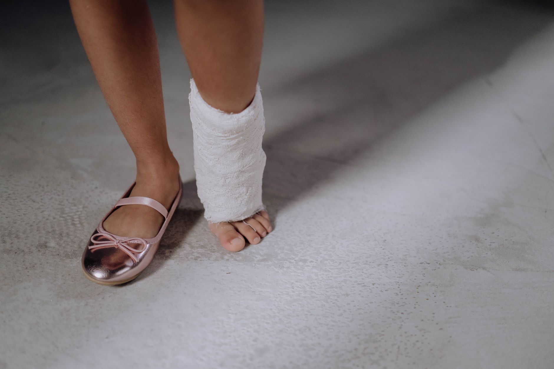
    RBD symptoms typically emerge in the 50s, 60s, or 70s, though earlier onset is possible. The most common presentation is a bed partner noticing kicking, talking, or yelling during sleep rather than the patient spontaneously reporting the behavior. Episodes occur multiple times per night, several nights a week, and often intensify over time. A person might repeatedly punch the air, shout obscenities during quiet dreams, or even fall off the bed, leading to bruises, cuts, or broken bones. One well-documented case involved a man who repeatedly dove headfirst onto the floor after dreaming he was jumping off a cliff—his partner eventually placed a mattress on the bedroom floor to prevent injury.

    The timing relationship between RBD onset and later Parkinson’s diagnosis varies widely. In some cases, RBD precedes motor symptoms by decades; in others, people develop mild RBD while already experiencing subtle motor changes they haven’t yet attributed to Parkinson’s. A few individuals are diagnosed with both RBD and Parkinson’s within months, suggesting rapid neurodegeneration. The average time from RBD diagnosis to Parkinson’s diagnosis is typically reported as 10 to 15 years in research cohorts, but this represents a median—some progress much faster, and others remain RBD-only for far longer. This unpredictability makes prognosis conversations difficult and underscores why regular neurological follow-up is essential.

    Sleep Study Findings and Risk Stratification

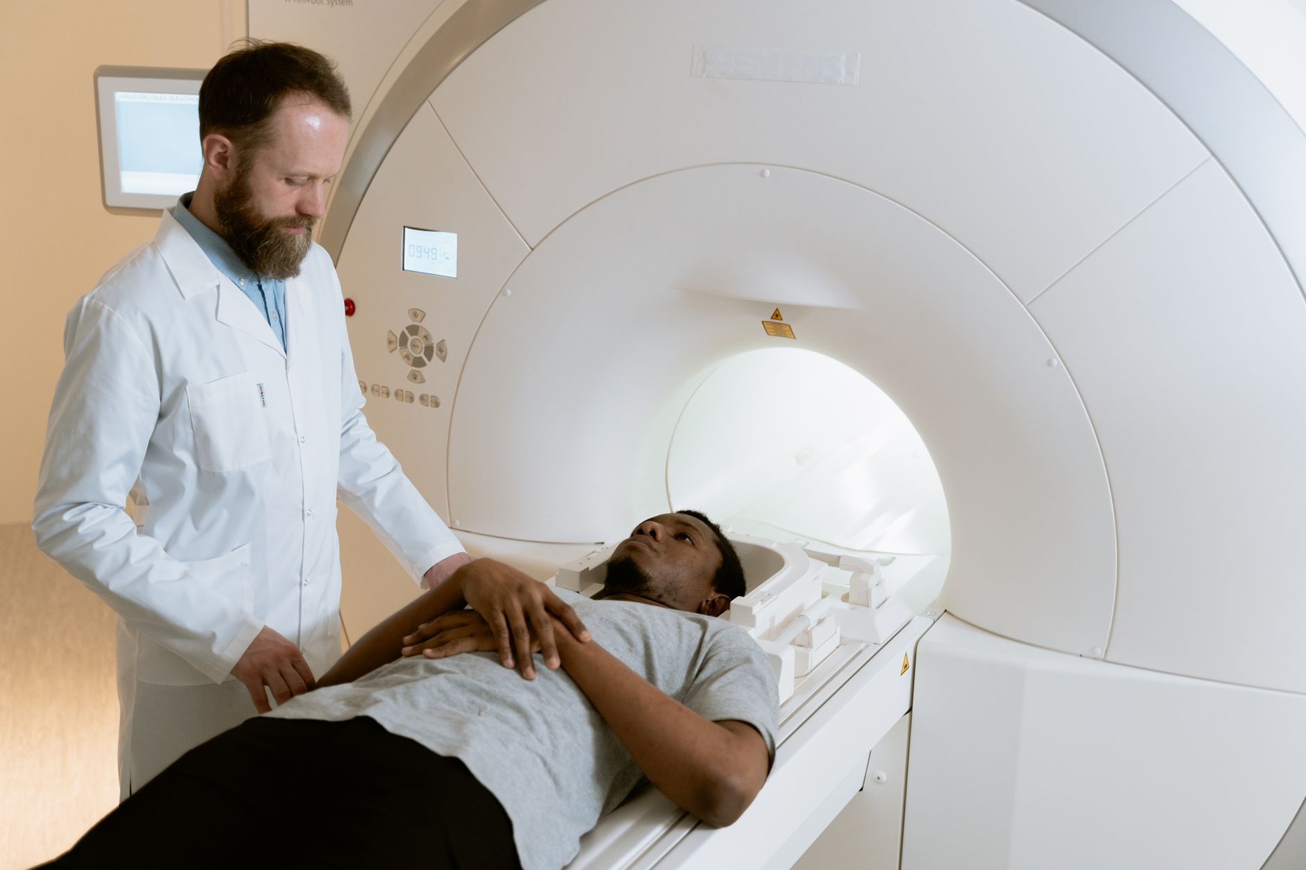
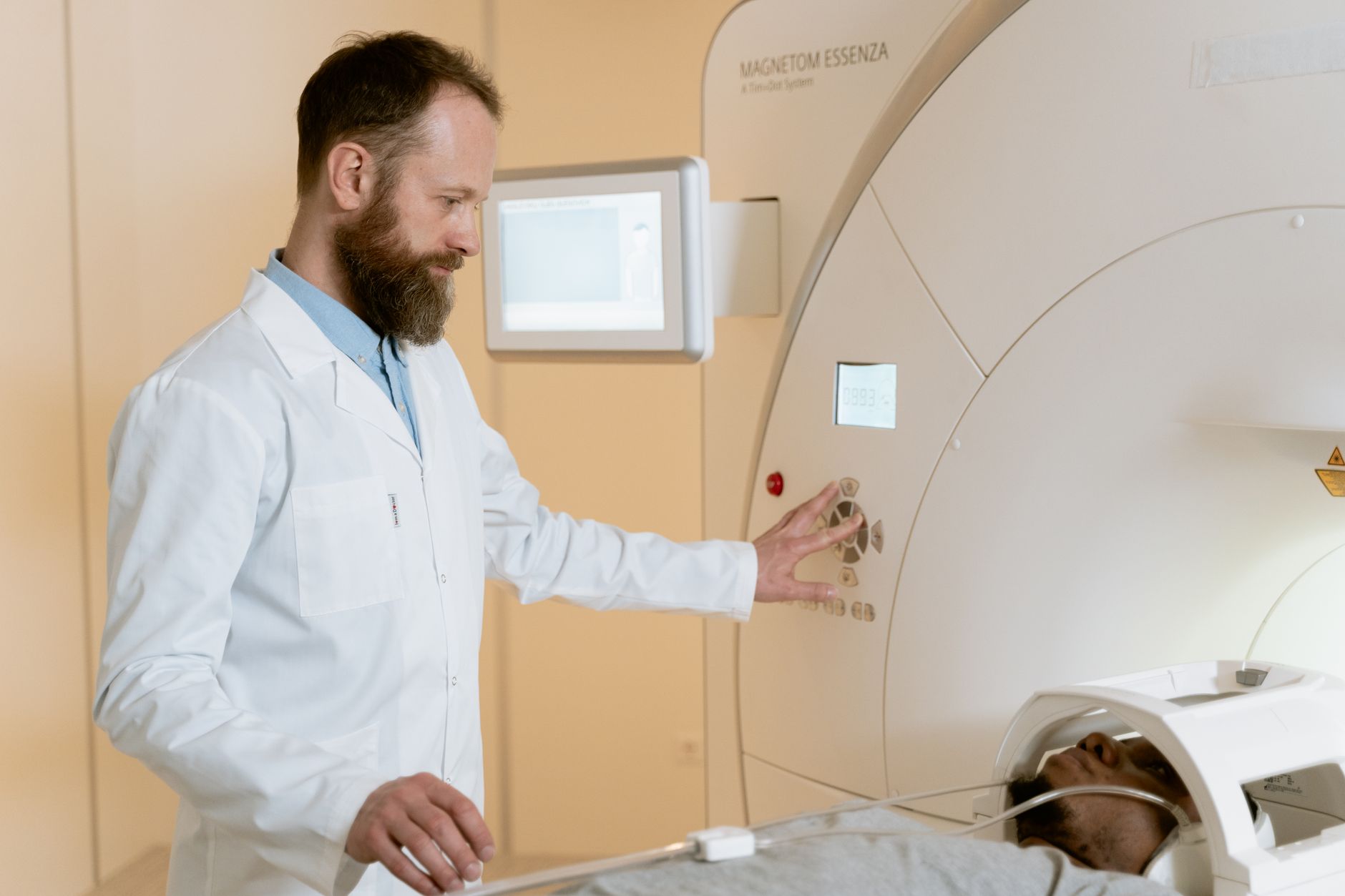
    A sleep study for suspected RBD involves an overnight polysomnographic recording, typically in a monitored lab setting where video surveillance captures movements and safety is ensured. The study quantifies REM sleep muscle tone and documents limb movements, providing objective confirmation beyond behavioral history alone. Some sleep centers perform video-PSG specifically designed to capture complex movements, which strengthens the clinical correlation. Risk stratification can then occur based on additional imaging or biomarker testing—quantitative motor assessment, dopamine transporter imaging (DaT scan), and cerebrospinal fluid alpha-synuclein levels may help predict progression likelihood, though these are not yet part of standard clinical practice for all RBD patients.

    A tradeoff exists between early identification and over-medicalization. Some patients, once diagnosed with RBD and told of the Parkinson’s link, experience significant anxiety that affects their own sleep quality and quality of life. Others find the diagnosis provides relief (explaining long-standing sleep disturbance) and motivation for lifestyle modifications. Sleep medications like melatonin (1-3 mg at bedtime) or low-dose clonazepam (0.25-1 mg at night) can reduce RBD episodes effectively, but these do not reverse the underlying neurodegeneration or prevent Parkinson’s progression—they simply suppress dream-acting behavior. The question of whether treating RBD with medications delays Parkinson’s onset remains unanswered and is an active area of research.

    Complications and Safety Considerations

    Injuries from RBD can be serious and sometimes tragic. Patients have broken ribs by striking bedside furniture, dislocated shoulders while thrashing, and sustained head injuries from falling. One documented case involved a 72-year-old man who sustained a subdural hematoma after hitting his head during a violent dream; another involved a woman who ran through a glass door during an episode. These aren’t rare outlier incidents—approximately 30 percent of RBD patients report at least one significant injury, and many have multiple minor injuries from repeated episodes.

    A major limitation in RBD research is the lack of consensus on when to screen for Parkinson’s aggressively or how often to monitor asymptomatic RBD patients; guidelines are evolving but remain heterogeneous across neurology practices. Sleep disruption itself becomes a problem. Night awakenings, the stress of episode-related injuries, and medication side effects can degrade sleep quality and increase daytime somnolence, which ironically may accelerate cognitive or motor decline in the context of incipient Parkinson’s. Depression and anxiety are common comorbidities in RBD patients, both independently associated with worse Parkinson’s outcomes. Some patients opt for environmental modifications—padding the bedroom, moving the mattress to the floor, using a body pillow barrier, or in severe cases, sleeping in separate beds from partners—which reduce injury risk but can strain relationships and social functioning.

    Neuropathological Evidence and Autopsy Studies

    Autopsy studies of people who had RBD during life have consistently shown alpha-synuclein pathology (Lewy bodies) in brainstem nuclei, supporting the hypothesis that RBD reflects early synucleinopathy. A landmark study of RBD patients who died revealed Lewy body pathology in 100 percent of examined brains, with a specific distribution pattern in the locus coeruleus and other REM-related structures. These findings confirm that RBD is not simply a sleep behavior problem but a manifestation of the same underlying neurodegeneration that characterizes Parkinson’s disease and Lewy body dementia.

    Screening, Follow-Up, and Current Clinical Practice

    Neurology and sleep medicine specialists increasingly incorporate screening for RBD history into appointments with older patients, particularly those with motor symptoms or family history of Parkinson’s. The RBD Screening Questionnaire (RBDSQ) is a validated tool that can prompt a polysomnography referral in primary care.

    Once RBD is diagnosed, recommended follow-up typically includes annual or biennial neurology visits with formal motor assessment (using scales like the Unified Parkinson’s Disease Rating Scale motor section) to detect emergence of Parkinson’s symptoms as early as possible. Some specialized centers offer longitudinal cognitive screening and dopamine transporter imaging to provide more granular risk prediction, though this is not yet standard of care everywhere. Genetic counseling may be relevant for patients with early-onset RBD or strong family history, as rare mutations (LRRK2, GBA) associated with Parkinson’s can also predispose to RBD.


    You Might Also Like

  • Can Constipation Be an Early Sign of Parkinson’s?

    Can Constipation Be an Early Sign of Parkinson’s?

    Yes, constipation can be an early sign of Parkinson’s disease. For many people with Parkinson’s, bowel problems appear years before the tremor, stiffness, or slowness of movement that most people associate with the condition. Research shows that constipation and other digestive changes affect up to 80 percent of people with Parkinson’s at some point during their disease, and for a significant portion, these gastrointestinal symptoms emerge in the decade before a Parkinson’s diagnosis is made.

    A 60-year-old man who suddenly began experiencing persistent constipation despite no dietary changes, combined with mild sleep disruption and reduced sense of smell, discovered three years later that these were the earliest markers of Parkinson’s disease. The connection between constipation and Parkinson’s stems from damage to nerve cells in the enteric nervous system—the network of nerves that controls the digestive tract. Because Parkinson’s affects the same type of nerve cells throughout the body, not just in the brain, the gut can show signs of neurological change long before motor symptoms become apparent. This is why doctors and caregivers should take persistent, unexplained constipation seriously, especially when it appears alongside other non-motor changes like mood shifts, sleep problems, or loss of smell.

    Table of Contents

    Is Constipation an Early Symptom of Parkinson’s Disease?

    Constipation is not universally an early sign of Parkinson’s—timing varies significantly from person to person. Some people experience bowel problems decades before other symptoms, while others develop constipation only after diagnosis when medications like dopamine agonists make it worse. Studies of newly diagnosed patients find that roughly 24 to 63 percent report having had constipation before their Parkinson’s diagnosis, which makes it more common than tremor as a first symptom, though less noticeable or concerning to most people.

    The challenge in recognizing constipation as an early warning is that it is extremely common in the general population and has many causes—diet, dehydration, aging, lack of physical activity, and dozens of medications can all contribute. What sets Parkinson’s-related constipation apart is its pattern: it typically develops gradually over months, does not fully resolve with standard remedies like increased fiber or water intake, and often coexists with other non-motor symptoms. A woman who had been managing mild constipation with over-the-counter stool softeners for five years, then added laxative dependency, tremors in her hand, and difficulty speaking to her list of complaints, was eventually diagnosed with Parkinson’s—and her gastroenterologist confirmed that the original constipation had likely been the disease’s first foothold.

    The enteric nervous system, sometimes called the “second brain,” contains roughly 500 million neurons that regulate digestion, movement, and secretions throughout the gastrointestinal tract. Parkinson’s disease damages dopamine-producing neurons, but the pathology is not limited to the brain—the same misfolded alpha-synuclein proteins that accumulate in the substantia nigra (the brain region responsible for movement control) also accumulate in the nerves of the gut, often in the earliest stages of disease. This means the digestive system can be affected before the motor circuits in the brain show significant damage. When these nerve cells in the gut are damaged, muscle contractions in the colon become uncoordinated and weak.

    The normal wave-like movements called peristalsis, which push stool through the intestines, slow down or become less effective. Additionally, Parkinson’s pathology can affect the autonomic nervous system, which controls involuntary functions like digestion and blood pressure regulation. A key limitation is that constipation from Parkinson’s is difficult to distinguish from constipation due to aging, diet, or other medical conditions without more definitive diagnostic markers. Doctors cannot diagnose Parkinson’s by constipation alone; the diagnosis requires the presence of motor symptoms or specific patterns of non-motor symptoms alongside a clinical assessment.

    Prevalence of Constipation Before Parkinson’s DiagnosisBefore Diagnosis32%At Diagnosis28%Within 5 Years of Diagnosis18%Develops Later15%Never Reported7%Source: Aggregated data from multiple Parkinson’s longitudinal and retrospective studies

    Timeline and Severity of Bowel Changes Before Motor Symptoms Appear

    The timing of constipation relative to a Parkinson’s diagnosis varies widely. In some people, bowel changes emerge 10 to 20 years before tremor or rigidity becomes noticeable. In others, constipation develops around the same time as motor symptoms or even afterward. Population studies suggest that among people who later develop Parkinson’s, those with chronic constipation in midlife have a higher statistical risk, but constipation in younger people is rarely a Parkinson’s precursor.

    The severity of constipation also does not predict the severity of Parkinson’s motor symptoms. Someone with mild, occasional constipation managed with dietary changes might eventually develop severe Parkinson’s, while another person with debilitating constipation requiring multiple medications might have slower disease progression. A man in his early 50s who experienced severe constipation requiring prescription laxatives was diagnosed with early-stage Parkinson’s with minimal tremor; conversely, a woman with very mild constipation discovered at routine exam years into her Parkinson’s journey found that her motor symptoms were more pronounced than her gastrointestinal ones. This unpredictability makes it difficult for patients or doctors to assume bowel problems are a reliable harbinger of motor disease.

    What to Do if You Notice Constipation in Yourself or a Loved One

    If constipation appears suddenly in an adult with no prior history of bowel problems, or if it persists despite reasonable dietary and lifestyle adjustments, it warrants a conversation with a primary care doctor. The doctor should ask about other non-motor symptoms: changes in smell, mood, sleep quality, energy level, or subtle motor changes like slower handwriting or reduced arm swing while walking. A record of when symptoms started can help establish a timeline.

    For someone already diagnosed with Parkinson’s, constipation management becomes more complex because Parkinson’s medications can worsen bowel function. Increasing dietary fiber and water without also increasing physical activity often makes Parkinson’s-related constipation worse, not better, because the underlying issue is nerve and muscle dysfunction, not just insufficient bulk in the diet. Working with a gastroenterologist familiar with Parkinson’s is valuable; they can recommend targeted interventions like osmotic laxatives, stool softeners, or medications that enhance gut motility in ways that differ from standard constipation treatment. The tradeoff is that many effective constipation medications have their own side effects or risks—some slow gastric emptying, others can interact with Parkinson’s drugs, and long-term laxative use can reduce the colon’s natural responsiveness.

    Other Non-Motor Symptoms That Often Appear with Constipation

    Constipation rarely appears in isolation as an early Parkinson’s symptom. Most people with Parkinson’s report a cluster of non-motor changes in the years before diagnosis: loss of smell (hyposmia), rapid eye movement sleep behavior disorder (acting out dreams), mood changes, anxiety, or cognitive shifts. Loss of smell is particularly notable because it is one of the earliest pathological changes in Parkinson’s and affects the olfactory bulb—a part of the brain supplied by similar neural circuits as the gut. When constipation appears alongside loss of smell and sleep disturbances, the statistical likelihood of Parkinson’s disease increases substantially.

    A warning: many of these non-motor symptoms are also common with aging, depression, or other neurological conditions. Someone experiencing constipation plus reduced sense of smell might have chronic sinusitis, medication side effects, or normal age-related change rather than Parkinson’s. Doctors sometimes dismiss these early signs as separate, unrelated issues—a patient complains of constipation to their primary care doctor, mentions smell loss to an ear-nose-throat specialist, and reports sleep problems to a sleep clinic, with no one seeing the pattern. This fragmentation of care can delay Parkinson’s recognition by years.

    The Role of Parkinson’s Medications in Worsening Constipation

    Once a Parkinson’s diagnosis is confirmed and medication begins, constipation often worsens. Dopamine agonists and other Parkinson’s drugs can slow intestinal movement further, and anticholinergic medications used to manage tremor directly impair digestive function by blocking acetylcholine, a neurotransmitter essential for muscle contractions in the gut. A person who had mild, manageable constipation before starting levodopa might find that within weeks of beginning treatment, bowel function becomes a significant daily burden.

    This creates a clinical dilemma: the medications that help control motor symptoms can make the very symptom that may have first signaled disease worse. Some patients and doctors respond by adding laxatives or stool softeners to the medication regimen, but this adds another layer of pharmacological management and potential interactions. An alternative approach involves emphasizing physical activity and abdominal massage, which can improve gut motility without additional drugs, though these approaches require consistent effort and are not always sufficient.

    Dietary and Lifestyle Strategies When Constipation May Signal Parkinson’s

    If constipation appears to be linked to early Parkinson’s—or even if there is uncertainty about its cause—lifestyle modifications should be attempted before or alongside medication. Adequate hydration, regular physical activity including walking or cycling, and structured abdominal massage can help stimulate bowel movements. However, the standard advice of “eat more fiber” often backfires in Parkinson’s because insufficient fluid intake, reduced physical activity, or existing nerve damage means the additional bulk has nowhere to go and can actually worsen symptoms.

    Specific timing matters: some people with Parkinson’s-related constipation benefit from warm liquids first thing in the morning to trigger the gastrocolic reflex, or from movement and activity immediately after meals. A person managing Parkinson’s-related constipation found that a 20-minute walk after breakfast, combined with warm coffee and adequate water throughout the day, was more effective than stool softeners alone. However, individual responses vary—what works for one person may not help another, and over time, the same strategy may become less effective as the disease progresses and nerve damage increases.


    You Might Also Like

  • Why Loss of Smell Can Appear Before Parkinson’s Movement Problems

    Why Loss of Smell Can Appear Before Parkinson’s Movement Problems

    Loss of smell often appears years or even decades before the tremor, rigidity, or slowness that defines Parkinson’s disease—a timing gap that puzzles many patients and families. The olfactory nerve and certain brain structures involved in smell degenerate early in Parkinson’s progression because of how the disease’s hallmark protein, alpha-synuclein, spreads through the nervous system. A person might notice they can no longer smell coffee or detect spoiled milk, yet move and function normally, all while changes are silently reshaping their brain in ways that will eventually affect movement control.

    This early appearance of smell loss exists because the olfactory bulb—a brain structure directly exposed to the environment through the nose—becomes one of the first targets of pathological alpha-synuclein accumulation. Some researchers theorize the protein may even enter the brain through olfactory neurons, making the nose a potential entry point for the disease process itself. For someone experiencing unexplained anosmia, or loss of smell, this symptom can be one of the most meaningful early warning signs of Parkinson’s, even when a neurologist cannot yet detect any movement abnormalities.

    Table of Contents

    How Does Alpha-Synuclein Damage the Olfactory System Before Motor Symptoms Appear?

    In Parkinson’s disease, alpha-synuclein protein misfolds and accumulates into clumps called Lewy bodies, which damage and kill nerve cells throughout the brain. The olfactory bulb is unusual because its neurons are constantly replaced throughout life, yet they become some of the first cells to accumulate these toxic protein deposits. This early and intense pathology in smell-related structures occurs long before the substantia nigra—the movement control center most associated with Parkinson’s—reaches the threshold of cell death needed to trigger visible motor symptoms.

    The olfactory epithelium, the tissue lining your nasal cavity that detects smells, sits directly on neurons that project into the brain. These neurons are uniquely vulnerable because they lack a strong blood-brain barrier protection and are repeatedly exposed to environmental toxins, pollutants, and pathogens. A person may lose 30, 50, or even 70 percent of their sense of smell and have no idea why, while elsewhere in their brain the disease is progressing through regions that regulate movement, emotion, and thinking. Research shows that roughly 90 percent of Parkinson’s patients eventually develop anosmia, but it frequently appears as an isolated symptom years before diagnosis.

    The Role of Braak Staging and Spreading Pathology in Olfactory Dysfunction

    Neuropathologists have mapped how Parkinson’s pathology spreads through the brain in a predictable pattern called Braak staging, and the olfactory bulb appears in stages 1 and 2—the earliest phases—often alongside the dorsal motor nucleus of the vagus nerve in the brainstem. This staging helps explain why smell loss is so common and so early: the disease starts in these anatomically isolated regions before it reaches the midbrain structures required for smooth movement. However, this staging model is not perfectly linear in every patient; some people accumulate pathology in multiple brain regions simultaneously, making the progression unpredictable at the individual level. One significant limitation of using smell loss as a diagnostic marker is that many conditions cause anosmia without any relationship to Parkinson’s.

    Chronic rhinosinusitis, allergies, COVID-19, head injury, and normal aging all impair smell. A 65-year-old who has lost smell gradually over five years might have years of mold exposure in their basement, not Parkinson’s disease. Only when smell loss is paired with other subtle signs—difficulty moving the fingers quickly, reduced facial expression, a softer voice, or a hunched posture—does anosmia become meaningful as a prodromal warning. Doctors often miss this connection because patients themselves do not link these seemingly separate problems or because they normalize gradual smell loss as part of aging.

    Prevalence of Non-Motor Symptoms in Parkinson’s Disease (%)Smell Loss90%Constipation80%Sleep Problems75%Depression40%Cognitive Changes25%Source: Parkinson’s Foundation; based on longitudinal patient surveys

    Early Detection Through Smell Testing and Clinical Recognition

    Specialized smell identification tests, most commonly the University of Pennsylvania Smell Identification Test (UPSIT), can quantify olfactory loss objectively and have become part of research protocols aimed at identifying people in the prodromal phase of Parkinson’s—that window before movement symptoms appear. A patient who scores in the impaired range on the UPSIT alongside other prodromal features like REM sleep behavior disorder, mild constipation, or depression may warrant closer monitoring and earlier neurological evaluation. This approach has identified hundreds of people at high risk for future Parkinson’s diagnosis, allowing research teams to study disease progression before it becomes clinically apparent.

    In clinical practice, few neurologists routinely test smell in patients who report it has declined. The conversation typically goes something like: “When did you notice?” “A few years ago.” “Could be aging.” End of discussion. This represents a missed opportunity, because a patient who lost smell five years ago and now has early motor signs is likely in mid-stage disease, whereas someone caught during the smell-loss-only phase might benefit from disease-modifying therapies if they become available. Some specialized movement disorder clinics now include smell testing as part of their standard evaluation for patients with suspected Parkinson’s or at-risk family members.

    Why the Olfactory Bulb Degenerates While Movement Centers Are Still Largely Intact

    The olfactory bulb’s vulnerability stems from its direct exposure to the external environment and its high metabolic demands. Unlike most brain structures protected by the blood-brain barrier, olfactory neurons extend directly from the nasal cavity through the cribriform plate—a thin bone separating the nose from the brain—into the olfactory bulb itself. This anatomy makes these cells exceptionally accessible to viruses, pollutants, and whatever pathological proteins might be traveling up from the gut through the vagus nerve, a theory gaining support in recent research. The substantia nigra, by contrast, is buried deep within the brain and surrounded by protective structures, so it takes longer for pathology to accumulate there to disease-causing levels.

    The tradeoff is significant: being early to degenerate makes the olfactory system a potential diagnostic window, but anosmia is entirely non-specific to Parkinson’s. A person losing smell should not assume they are developing Parkinson’s, as the vast majority of smell loss has other causes. Neurologists must weigh whether smell loss alone, without other prodromal features or family history, warrants preventive monitoring or whether it simply reflects common conditions like sinusitis or post-viral dysfunction. For someone with multiple prodromal markers—smell loss plus REM sleep behavior disorder plus autonomic symptoms—the picture becomes clearer and more concerning for future Parkinson’s risk.

    Prodromal Parkinson’s and the Importance of Recognizing Subtle Multi-System Changes

    Prodromal Parkinson’s refers to the period when alpha-synuclein pathology is accumulating and early non-motor symptoms appear, but movement dysfunction is not yet obvious enough for formal diagnosis. Alongside smell loss, people in this phase often report constipation, sleep problems, mood changes, or autonomic symptoms like blood pressure fluctuations—all potentially related to alpha-synuclein spread through the brainstem and peripheral nervous system. A person might see their primary care doctor for constipation, then their sleep specialist for insomnia, then an ENT for anosmia, without anyone recognizing these as part of a unified disease process. A critical warning here is that people identified as prodromal do not inevitably develop motor Parkinson’s on any particular timeline.

    Some people with smell loss, REM sleep behavior disorder, and other prodromal markers remain in that phase for 5, 10, or even 20 years without developing tremor or rigidity. Others progress rapidly. Research is still determining which prodromal features predict faster progression and which protective factors might slow or prevent the transition to clinical Parkinson’s. Current medical advice focuses on monitoring, exercise, sleep optimization, and cardiovascular health rather than starting Parkinson’s medications in asymptomatic people, even when prodromal signs are evident.

    The Potential Gateway Hypothesis—Smell, the Vagus Nerve, and Ascending Pathology

    Some neuroscientists propose that Parkinson’s pathology may enter the brain through the olfactory nerve or through the vagus nerve in the gut, then spread upward to affect progressively higher brain structures. Evidence for this “gateway” or “body-first” hypothesis includes the strong connection between olfactory loss and future Parkinson’s, the early involvement of the dorsal motor nucleus of the vagus nerve in brainstem pathology, and the commonality of gastrointestinal symptoms in Parkinson’s.

    If this theory holds true, understanding why the olfactory system is compromised early might unlock clues to disease initiation and offer new prevention or early-stage intervention targets. This hypothesis remains speculative, and human studies cannot yet directly prove that alpha-synuclein travels from nose to brain or from gut to brain in Parkinson’s patients. Animal studies have shown that engineered alpha-synuclein can travel along neural pathways, and some epidemiological studies have linked viral infections and gut dysbiosis to later Parkinson’s risk, but the chain of causation in humans is not fully established.

    Implications for Current and Future Diagnostic and Monitoring Approaches

    Olfactory testing is already being incorporated into research protocols designed to identify and monitor people at high genetic or phenotypic risk for Parkinson’s. Longitudinal studies following people with smell loss and other prodromal features are underway in multiple countries, creating databases that will eventually clarify which combinations of prodromal markers best predict future diagnosis and progression speed.

    For patients and families with Parkinson’s history or unexplained anosmia, participating in such research can provide valuable follow-up monitoring and contribute to understanding disease origins. In the clinical setting, a person who notices they cannot smell their favorite foods or has repeatedly been surprised by spoiled groceries should mention this to their physician, especially if they have a family history of Parkinson’s or are over age 50. The loss of smell alone is unlikely to warrant extensive neurological workup, but combined with other subtle signs—constipation, tremor, slow movements, reduced arm swing, soft voice, or sleep disturbances—it becomes part of a pattern worth investigating with a neurologist experienced in movement disorders.


    You Might Also Like

  • What Is Usually the First Noticeable Sign of Parkinson’s?

    What Is Usually the First Noticeable Sign of Parkinson’s?

    The most commonly reported first sign of Parkinson’s disease is a tremor—usually a resting tremor that starts in one hand and occurs when the limb is at rest. Many people notice a subtle shaking in their fingers or hand when sitting or relaxing, which may intensify under stress and disappear during intentional movement. This tremor affects roughly 70% of people at disease onset, making it the single most recognizable early symptom, though not everyone experiences it.

    However, a significant minority of Parkinson’s patients—approximately 25 to 30%—develop the disease without any tremor at all. For these individuals, the first noticeable sign might be slowness of movement (bradykinesia), stiffness in the limbs, difficulty with balance, or subtle changes in handwriting. The variability in early presentation means that some people delay seeking diagnosis because they don’t experience the tremor they expected, or because their initial symptoms seem minor or unrelated to a neurological condition.

    Table of Contents

    How Does Tremor Typically Present as a First Sign?

    When tremor is the first sign, it usually manifests as a low-frequency oscillation—typically 4 to 6 cycles per second—that occurs primarily when the hand or arm is at rest and supported. A person might notice their thumb and forefinger moving in a rhythmic “pill-rolling” motion, or a general shaking in their palm when their hand rests on a table or in their lap. The tremor often appears unilaterally first, affecting one side of the body before potentially progressing to the other side over months or years. Importantly, Parkinsonian tremor diminishes or disappears when the person actively uses the affected limb. Someone with an early tremor in their right hand might write, eat, or gesture without any apparent shaking, but the tremor returns immediately when the hand rests.

    This distinguishes it from essential tremor, which worsens with intentional movement—a key difference that helps clinicians differentiate between the two conditions. A person might first notice their tremor while watching television, during a car ride, or in meetings where their hand is still. Stress, fatigue, and cold temperatures can exacerbate Parkinsonian tremor, sometimes making it dramatically more obvious. A patient might report that their tremor is barely noticeable on a relaxed morning but becomes pronounced by late afternoon, or that it worsens noticeably during a stressful work presentation. This variability can lead to delayed diagnosis if the tremor is intermittent or context-dependent, and the person doesn’t consistently observe it during medical appointments.

    Why Isn’t Tremor Always the First Sign?

    In akinetic-rigid Parkinson’s disease—a subtype that accounts for 15 to 25% of cases—tremor is absent or minimal from onset, and instead slowness and stiffness dominate early presentation. These patients experience bradykinesia (slowed movement) and rigidity that might first appear as difficulty rising from a chair, challenges with fine motor tasks like buttoning clothes, or a general sense of motor fatigue. Without the obvious tremor, these individuals may attribute their symptoms to aging, arthritis, or depression rather than seeking neurological evaluation. Other non-tremor early signs include postural instability (balance problems), gait changes (shorter steps, reduced arm swing when walking), voice changes (softer, more monotone speech), or micrographia (progressively smaller handwriting).

    Some people notice reduced facial expression or a fixed expression before any motor slowness becomes apparent. The challenge with these presentations is that none of them are specific to Parkinson’s—balance problems could suggest inner ear issues, soft speech might be attributed to fatigue, and small handwriting could simply reflect aging. A limitation in relying on tremor as the diagnostic indicator is that many conditions produce similar tremors or movement abnormalities, and primary care physicians may initially misdiagnose the condition as essential tremor, hyperthyroidism, anxiety disorder, or Alzheimer’s disease. Patients presenting without tremor face an even higher risk of delayed diagnosis, as the early symptoms are more subtle and easily confused with other age-related changes or neuropsychiatric conditions.

    Percentage of Parkinson’s Patients by Initial Symptom TypeTremor-Dominant70%Rigidity/Bradykinesia-Dominant20%Postural Instability-Dominant5%Mixed Presentation3%Non-Motor Signs Only2%Source: Movement Disorder Society Clinical Guidelines; Parkinson’s Foundation

    Variations in How Symptoms Develop Across Individuals

    Two people with Parkinson’s disease can have entirely different first symptoms and follow different disease trajectories. One 62-year-old woman might notice that her right hand tremors when relaxed, while her 65-year-old neighbor develops progressive stiffness in his shoulders and neck without any tremor for several years. These variations are not fully understood but appear related to where neurodegeneration begins in the brain and how it spreads, as well as individual genetic and environmental factors. The side of the body where symptoms first appear often remains the more severely affected side throughout the disease course, a phenomenon called “motor asymmetry.” Someone whose tremor begins on the right side may find that right-sided symptoms remain more prominent even decades into the disease.

    This asymmetrical pattern can be helpful for diagnosis but also means that early presentation gives clues about the likely pattern of long-term progression for that individual. Age at onset also influences the nature of first symptoms. Younger-onset Parkinson’s patients (before age 50) are statistically less likely to present with tremor and more likely to first notice rigidity, bradykinesia, or dystonia (involuntary muscle contractions). Older patients have a higher rate of tremor-dominant presentation. This age-related difference suggests that Parkinson’s disease may have subtly different underlying mechanisms depending on when neurodegeneration begins, which has implications for how quickly the disease progresses and how it responds to treatment.

    Recognizing Early Signs and Deciding When to Seek Evaluation

    The challenge for most people is distinguishing a genuinely abnormal first sign from normal aging or stress-related symptoms. A person who experiences occasional hand shaking might reasonably wonder whether this is Parkinsonian tremor or simply anxiety-related tremor or caffeine sensitivity. A useful distinction is that Parkinsonian tremor is persistent over weeks and months, occurs at rest without clear external triggers, and often has a regular, rhythmic quality. When other early signs appear alongside tremor or instead of it, the case for seeking medical evaluation becomes stronger.

    Difficulty with handwriting, reduced arm swing while walking, difficulty turning over in bed, trouble with facial expression, or unexpected fatigue during routine activities warrant a neurology consultation. A person should not ignore these changes simply because they seem minor—Parkinson’s disease is often diagnosed when early motor signs are still subtle, and early diagnosis offers access to symptomatic treatments and potentially disease-modifying therapies under development. Primary care physicians can perform basic screening tests like asking about tremor at rest, observing gait and posture, and assessing bradykinesia with simple tasks like rapid finger tapping or hand pronation-supination tests. However, a neurology referral is advisable for definitive diagnosis, especially when the clinical presentation is atypical or when symptoms could suggest alternative diagnoses. There is no downside to earlier evaluation, as early diagnosis allows patients to plan their lives, optimize their treatment, and potentially participate in clinical trials.

    Delayed Diagnosis and Why Early Signs Are Often Missed

    Many people live with undiagnosed Parkinson’s disease for months or even years before seeking or receiving a diagnosis. Some delay going to a doctor because they attribute symptoms to normal aging, stress, or minor health issues. Others visit physicians who do not recognize early Parkinsonian features or misattribute them to other conditions—a primary care doctor might diagnose “essential tremor” without further neurological testing, or attribute bradykinesia and fatigue to depression. The risk of misdiagnosis is particularly high in patients without tremor, who may be told they have “normal aging,” arthritis, or psychological conditions before a neurologist eventually recognizes the pattern.

    A 70-year-old with progressive stiffness and slow movement might spend years in physical therapy for presumed arthritis before a neurologist diagnoses Parkinson’s disease. This delay means lost opportunity for early symptomatic treatment and potential exclusion from clinical trials that target early-stage disease. Cognitive bias also plays a role—if a person doesn’t know that non-tremor presentations of Parkinson’s exist, they may not recognize their own symptoms as potentially neurological. Public awareness of Parkinson’s disease tends to emphasize tremor, leaving many people unaware that stiffness, balance problems, or voice changes can be equally significant early signs. This underscores the importance of education not just for patients but for healthcare providers about the full spectrum of early Parkinson’s presentations.

    The Role of Family History and Genetic Risk

    Approximately 10 to 15% of people with Parkinson’s disease have a family history of the condition, and specific genetic mutations increase disease risk. Someone with a parent or sibling with Parkinson’s should be alert to early signs and may want to establish a baseline neurology evaluation even if symptoms haven’t appeared. Genetic testing is available for some Parkinson’s-associated mutations, though the presence of a mutation does not guarantee disease development, and most people with Parkinson’s have no genetic mutation that has been identified.

    For those with genetic risk, early recognition of first signs becomes even more important. A family member with known disease might recognize subtle symptoms in a relative earlier than they would otherwise appear on medical radar. However, genetic information also carries psychological weight—someone who knows they carry a Parkinson’s-linked mutation but has not yet developed symptoms may experience anxiety about whether and when symptoms will emerge. This highlights the importance of discussing both the benefits and limitations of genetic testing with a neurologist.

    Early Symptom Documentation and the Importance of Detailed History

    When someone suspects they may have early Parkinson’s disease, keeping a detailed record of when symptoms started, how they’ve changed, and what triggers them can be invaluable for a neurologist’s evaluation. Noting when tremor first appeared, whether it was constant or intermittent, whether it worsened over weeks or months, and how it responds to movement and stress provides concrete clinical information. Similarly, documenting changes in handwriting, gait, or speech over a timeline helps establish whether changes are progressive and consistent with neurological disease.

    A patient might photograph or record video of tremor or other symptoms to show their neurologist, especially if symptoms are intermittent or less obvious in the office setting. Keeping a brief symptom diary for a few weeks before a neurology appointment—noting when tremor or other signs are most noticeable and under what circumstances—gives the physician a clearer picture of the pattern. This level of detail has improved diagnostic accuracy and helps distinguish Parkinson’s disease from other conditions that can mimic early Parkinsonian signs.


    You Might Also Like

  • Can Parkinson’s Disease Begin Without a Tremor?

    Can Parkinson’s Disease Begin Without a Tremor?

    Yes, Parkinson’s disease can absolutely begin without a tremor. In fact, research shows that tremor is absent at initial diagnosis in 25 to 50 percent of people with Parkinson’s. A 52-year-old woman might notice her right leg feels stiff during morning walks, or her arm moves awkwardly when she reaches for things, only to learn months later—after struggling with these symptoms—that she has Parkinson’s.

    The absence of the classic “pill-rolling” tremor doesn’t mean the disease isn’t present or progressing. Many people expect Parkinson’s to announce itself with the visible shaking they’ve seen in films or news stories. But the condition can creep in through rigidity, slowness of movement, balance problems, or difficulty with fine motor tasks. These non-tremor symptoms are just as much a part of Parkinson’s disease as the shaking people associate with the diagnosis.

    Table of Contents

    What Non-Tremor Parkinson’s Symptoms Actually Feel Like

    rigidity—a stiffness in the muscles that makes movement feel heavy or resistant—is often the first symptom someone with Parkinson’s experiences. This isn’t ordinary muscle tightness; it’s a constant resistance in the joints that worsens with movement and can feel like moving through thick mud. A man in his 60s might wake up and find his shoulder won’t rotate fully, or his neck feels locked when he tries to look over his shoulder. This rigidity can appear in one limb first, typically on one side of the body, and worsen over weeks or months. Bradykinesia, or slowness of movement, frequently appears alongside or instead of tremor.

    Everyday actions slow down—buttoning a shirt takes twice as long, handwriting becomes smaller and more laborious, or walking feels like each step requires conscious effort. One person might notice they’re moving much more slowly than they used to, or a spouse might point out that their facial expressions seem less animated, a symptom called “masked face” that happens because the muscles controlling expression move more slowly. These symptoms can be subtle enough that people dismiss them as signs of aging or stress. A woman might assume her small, cramped handwriting is just because she needs glasses, or a man might blame his slowness on not exercising enough. The insidious nature of non-tremor Parkinson’s is that it can advance without drawing attention to itself the way a visible tremor would.

    The Reality of Early Parkinson’s Symptoms

    Postural instability—difficulty maintaining balance and coordination—can emerge early and become a serious safety concern. Unlike tremor, which someone might notice themselves, balance problems often become apparent when a person nearly falls while turning, or when they take unsteady steps while walking. A 60-year-old woman living alone might have a fall she attributes to a loose rug, unaware it was an early sign of Parkinson’s affecting her balance and coordination. The limitation of relying on tremor as a key diagnostic marker is that patients without tremor often take longer to receive an accurate diagnosis.

    Instead of getting a Parkinson’s diagnosis within months of symptom onset, non-tremor patients might see neurologists who initially rule out Parkinson’s, get misdiagnosed with other conditions like depression or normal aging, and experience unnecessary delays in starting treatment. During this time, symptoms may worsen and people often develop anxiety about what’s happening to their body without a clear explanation. Sleep disturbances, including insomnia and restless sleep, can also be early signs in the absence of tremor. Some people with early Parkinson’s experience vivid, sometimes disturbing dreams, or wake up multiple times throughout the night. These symptoms might lead to a sleep specialist referral before anyone considers Parkinson’s disease, further delaying proper diagnosis and care.

    Presenting Motor Symptoms at Parkinson’s Diagnosis (Percentage of Patients)Tremor40%Rigidity70%Bradykinesia (Slowness)80%Balance Problems35%Combined45%Source: Movement Disorder Society clinical consensus data; percentages reflect overlap as multiple symptoms often present simultaneously

    Why Tremor Isn’t Always the First Sign

    The cause of tremor in Parkinson’s involves disrupted communication in specific brain circuits, but rigidity and slowness result from a somewhat different distribution of damage to dopamine-producing cells. Not everyone’s Parkinson’s develops in the same way. Some people inherit genetic variants that predispose them to non-tremor presentations, while others develop the disease through environmental or sporadic factors that affect different neural pathways. A person whose parent had Parkinson’s with prominent tremor might experience an entirely different symptom profile because the underlying genetic contribution differs.

    Age and gender also influence symptom presentation. Older adults and women are more likely to present with akinesia—the loss of automatic movement—rather than tremor. Young-onset Parkinson’s (before age 50) often presents differently than later-onset disease, sometimes with more rigidity and less tremor than typical. A woman diagnosed at age 45 with stiffness and slowness might have an entirely different trajectory than a man diagnosed at 75, yet both have Parkinson’s disease.

    Getting Diagnosed Without Tremor

    Diagnosis relies on clinical assessment rather than any single symptom. A neurologist looks for the constellation of motor symptoms—whether tremor is present or not—along with response to levodopa medication. If someone with rigidity and slowness who started dopamine replacement therapy experiences significant improvement in those symptoms, the diagnosis is confirmed even without tremor ever appearing. However, the diagnostic process can be more complicated when tremor isn’t the presenting feature.

    A significant tradeoff exists: tremor is easy to observe and document, while rigidity and slowness require skilled clinical examination. A doctor with limited experience in Parkinson’s disease might miss early signs of rigidity or confuse slowness with depression. This is why patients without tremor benefit from seeing a movement disorder specialist—a neurologist with specific expertise in Parkinson’s disease—rather than a general neurologist. Insurance and access barriers mean not everyone can see a specialist quickly, and some people spend considerable time being evaluated by physicians who aren’t attuned to non-tremor presentations.

    Common Misdiagnoses When Tremor Isn’t Present

    When a patient presents with rigidity, slowness, and balance problems but no tremor, physicians might initially suspect depression, fibromyalgia, arthritis, or normal aging. A 55-year-old woman who describes fatigue, slowness, and low mood might be started on an antidepressant and never evaluated for Parkinson’s. While depression does co-occur with Parkinson’s and should be treated, missing the Parkinson’s diagnosis means missing the opportunity for dopamine-replacement therapy that could address her motor symptoms directly. A critical warning: some people with non-tremor Parkinson’s have been told their symptoms are psychosomatic or stress-related, leading to unnecessary psychiatric treatment while the underlying neurological disease progresses untreated.

    Years can pass between symptom onset and accurate diagnosis, during which time the person may feel dismissed or blamed for their condition. The psychological toll of prolonged diagnostic uncertainty, combined with worsening physical symptoms, can create genuine depression—a secondary condition layered on top of the original neurological problem. Orthopedic misdiagnosis is also common. Rigidity in the neck and shoulders can mimic cervical arthritis, and someone might pursue physical therapy or even orthopedic surgery for a problem that would respond better to Parkinson’s medications. A man with rigidity might be told he has a frozen shoulder and prescribed weeks of physical therapy, only to discover the real cause was Parkinson’s disease.

    Living with Non-Tremor Parkinson’s

    Non-tremor Parkinson’s can feel more isolating because it lacks the visible hallmark people recognize. Friends and family might not immediately understand why someone is moving slowly or struggling with fine motor tasks, since there’s no visible shaking to point to as an explanation. A person might face skepticism about their symptoms or pressure to “just push through it,” not realizing that pushing through Parkinson’s symptoms often makes them worse, not better.

    The experience of rigidity in particular can be painful. Stiffness that resists movement generates physical discomfort, especially when someone tries to move against that resistance. Dopamine replacement therapy helps reduce rigidity, but the period before diagnosis—when someone is struggling with worsening stiffness without knowing why—can be marked by significant physical pain and frustration.

    How Movement Patterns Change Over Time

    Non-tremor Parkinson’s typically progresses through observable changes in walking, balance, and coordination. Someone who initially noticed shoulder stiffness might later find their gait narrowing—their stride becoming shorter, their steps quicker but less controlled—a pattern called festinating gait that increases fall risk significantly. Hand function may deteriorate to the point where writing, typing, or using utensils becomes frustratingly difficult long before tremor ever appears.

    A person with early-stage non-tremor Parkinson’s might maintain nearly normal function with medication and physical therapy for years, while another might experience faster progression. The variability in non-tremor presentations means that two people with identical diagnoses can have very different lived experiences. One person’s main challenge might be balance and fall prevention, while another struggles primarily with the slowness that makes self-care tasks take hours instead of minutes. Understanding your specific symptom pattern allows for targeted strategies: someone with rigidity benefits from regular stretching and resistance exercise, while someone with balance problems needs structured fall prevention and possibly assistive devices.


    You Might Also Like

  • How Parkinson’s Disease Affects Dopamine Production

    How Parkinson’s Disease Affects Dopamine Production

    Parkinson’s disease causes the progressive death of dopamine-producing cells in the brain, specifically in a region called the substantia nigra. This loss of dopamine—a critical neurotransmitter that coordinates movement, motivation, and emotional processing—is the primary reason Parkinson’s patients develop the disease’s hallmark symptoms of tremor, rigidity, and slowness of movement. For a person with early-stage Parkinson’s, dopamine levels may drop to 30 to 40 percent of normal; as the disease advances, this deficit can exceed 80 percent, fundamentally altering how the brain sends movement signals throughout the body.

    The cascade of neurological changes begins silently. Most people don’t notice symptoms until the loss of dopamine-producing neurons reaches a critical threshold—usually after roughly 50 to 60 percent of these cells have already died. This lag between cellular death and noticeable symptoms is why Parkinson’s is often diagnosed years after the underlying degeneration has started, a reality that makes understanding dopamine’s role essential for patients and caregivers who want to recognize early warning signs.

    Table of Contents

    What Is Dopamine and Why Does the Parkinson’s Brain Lose It?

    Dopamine is a neurotransmitter that acts as a chemical messenger between nerve cells. In a healthy brain, dopamine produced in the substantia nigra travels to other brain regions to coordinate smooth, intentional movement. It also plays a role in motivation, mood regulation, and the ability to initiate action—which is why dopamine deficiency affects far more than just motor control. A person with adequate dopamine can start walking across a room effortlessly; someone with Parkinson’s-related dopamine loss may need to consciously think through each step, and the action itself feels labored.

    In Parkinson’s disease, the cells that produce dopamine (called dopaminergic neurons) gradually deteriorate and die. Researchers have identified clues pointing to multiple causes: some neurons accumulate a protein called alpha-synuclein, others are damaged by oxidative stress, and still others fall victim to inflammation or genetic factors. The exact trigger remains unclear, which is why Parkinson’s is still considered idiopathic—arising from no single, well-understood cause. Unlike a stroke, where dopamine loss is sudden and localized, Parkinson’s involves a slow, region-specific erosion that leaves some dopamine-producing capacity intact even in advanced stages.

    How Dopamine Depletion Leads to Parkinson’s Symptoms

    When dopamine levels drop, the brain struggles to send the smooth, coordinated signals needed for movement. The substantia nigra normally maintains a delicate balance of chemical activity, and dopamine is central to that stability. Without sufficient dopamine, neural circuits that coordinate the timing and force of muscle contractions become imbalanced. The result is the characteristic tremor at rest, muscular rigidity (stiffness), and bradykinesia (slowness of movement) that define Parkinson’s.

    A critical limitation of current treatment is that while medications can increase dopamine availability in the brain, they cannot halt the underlying death of dopamine-producing neurons. This means that as more neurons die over time, even optimal medication becomes less effective. A 55-year-old diagnosed with Parkinson’s might respond well to levodopa for 5 to 7 years, then notice that the medication’s effect wears off more quickly each day, or that side effects become more pronounced. This phenomenon, called “motor fluctuations,” reveals the hard truth: dopamine replacement therapy treats the symptom but not the disease’s progressive nature.

    Dopamine Neuron Survival Across Parkinson’s Disease StagesHealthy Brain100%Early Parkinson’s50%Moderate Parkinson’s30%Advanced Parkinson’s15%Late-Stage Parkinson’s10%Source: Neurobiology of Parkinson’s Disease Research Synthesis

    Dopamine Loss and the Full Range of Parkinson’s Movement Problems

    Tremor, the visible shaking that many people associate with Parkinson’s, occurs because the loss of dopamine disrupts the brain’s ability to dampen unnecessary nerve impulses. However, not all Parkinson’s patients develop noticeable tremor—some experience rigidity and slowness as their primary symptoms. A patient might notice they can no longer button a shirt quickly, or that their handwriting has become very small, changes driven by the brain’s reduced dopamine-mediated control over fine motor tasks.

    Postural instability, a dangerous symptom that develops as dopamine deficiency worsens, arises because the brain’s sense of balance and righting reflexes depend on healthy dopamine signaling in multiple brain regions. Freezing of gait—an abrupt, involuntary halt while walking—also stems from dopamine loss affecting the areas of the brain responsible for automatic movement initiation. Someone with Parkinson’s might walk normally down a hallway, then freeze at a doorway, their feet feeling glued to the floor for seconds or minutes. This symptom does not respond reliably to dopamine medications, underscoring how the disease affects neural circuits beyond simple dopamine availability.

    Why Dopamine Medications Work Differently as the Disease Progresses

    Levodopa, the gold-standard medication for Parkinson’s, is converted to dopamine in the brain, temporarily replacing the dopamine lost to neuronal death. In early disease, when the brain still has intact dopamine-producing neurons, a dose of levodopa can be absorbed, stored, and released smoothly over several hours. The patient experiences steady symptom relief. However, as neuronal death accelerates and fewer dopamine-producing neurons remain, the brain loses its ability to store dopamine, making medication effects more unpredictable.

    By the mid to late stages of Parkinson’s, the same levodopa dose might work brilliantly for an hour, then wear off suddenly, leaving the patient either “on” (symptom-free) or “off” (severely symptomatic) with little middle ground. This creates a treatment dilemma: increasing the dose can intensify side effects like dyskinesia (involuntary, writhing movements), while lowering the dose leaves the patient with inadequate symptom control. A 70-year-old who has had Parkinson’s for 15 years often requires far more complex medication timing than a newly diagnosed patient, yet paradoxically experiences less consistent benefit. The tradeoff between symptom control and medication side effects becomes sharper as dopamine depletion advances.

    Non-Motor Symptoms and Hidden Dopamine Deficiency

    While movement problems dominate the public image of Parkinson’s, dopamine deficiency affects motivation, mood, and emotional regulation in ways patients and families often overlook until they become severe. Apathy—a loss of motivation and initiative separate from depression—affects roughly one-third to one-half of Parkinson’s patients and arises directly from dopamine loss in brain regions linked to reward and motivation. A person might lack the drive to bathe, socialize, or pursue hobbies despite having the physical ability to do so. This is not laziness or depression; it is dopamine deficiency dampening the internal signal that makes activities feel worth doing.

    Depression and anxiety also occur at higher rates in Parkinson’s than in the general population, partly because dopamine loss extends to brain circuits governing mood. A warning: depression and apathy in Parkinson’s sometimes fail to respond to standard antidepressants, because the underlying problem is dopamine deficiency, not serotonin shortage. Additionally, cognitive slowness—difficulty concentrating, retrieving words, or making decisions—reflects dopamine depletion in cortical regions involved in executive function. These non-motor symptoms are as disruptive to quality of life as tremor or rigidity, yet are frequently underrecognized and undertreated.

    Genetic and Environmental Clues to Dopamine Neuron Vulnerability

    Research into why certain people develop Parkinson’s has identified genetic mutations that affect dopamine system health. People carrying mutations in genes like LRRK2 or PINK1 have a higher risk of Parkinson’s, and these genes are involved in cellular functions that protect dopamine neurons.

    Environmental exposures, particularly to certain pesticides and herbicides, have been linked to dopamine neuron damage in epidemiological studies. A farmer exposed to paraquat or rotenone over decades faces a higher risk of Parkinson’s than someone with no agricultural chemical exposure, though exposure alone is not deterministic—genetics, age, and other factors also play a role.

    The Irreversibility of Dopamine Neuron Loss and What It Means for Treatment Strategy

    Unlike a neuron damaged by temporary toxin exposure or inflammation, a dopamine neuron killed by Parkinson’s disease does not regenerate. This irreversibility fundamentally shapes treatment: current medications manage symptoms by increasing dopamine activity in the remaining neurons, but they do nothing to replace the dead cells. No medication yet developed can restore dopamine production by reactivating dead neurons or prompting the brain to grow new dopamine-producing cells at scale. Clinical trials of neuroprotective agents—drugs designed to slow neuronal death—have shown mixed results, and no neuroprotective drug is currently approved for routine use in Parkinson’s.

    This means that treatment is inherently a race against progressive degeneration. A person diagnosed at age 60 with early-stage Parkinson’s is managing a condition where the underlying pathology continues even as medications provide symptom relief. The rate of dopamine neuron loss varies from person to person—some patients’ disease progresses slowly over 20 years, while others see faster decline. A patient whose dopamine neurons are dying at a rate of 5 percent per year will require increasingly complex medication management than someone whose decline is 2 percent per year, yet current diagnostic tools cannot accurately predict individual progression rates at the time of diagnosis.


    You Might Also Like

  • Who Is Most Likely to Develop Parkinson’s Disease?

    Who Is Most Likely to Develop Parkinson’s Disease?

    Parkinson’s disease is most likely to develop in adults aged 60 and older, with men diagnosed approximately 1.5 times more frequently than women. While age remains the single strongest risk factor, the actual likelihood of developing Parkinson’s involves a complex combination of genetics, environmental exposures, and individual health history that varies significantly from person to person.

    The majority of people diagnosed with Parkinson’s—about 90 percent—first show symptoms after age 50. However, younger adults can develop the condition too; roughly 5 to 10 percent of cases occur before age 40, sometimes called early-onset Parkinson’s disease. A 65-year-old man with a parent who had Parkinson’s faces a notably higher risk than a 65-year-old woman with no family history, illustrating how multiple factors interact to influence who develops the disease.

    Table of Contents

    Why Age Matters Most in Parkinson’s Development

    Age is the most reliable predictor of Parkinson’s disease risk, with the incidence increasing dramatically after age 60. The condition is rare before age 40 but becomes increasingly common with each decade of life. By age 80, approximately 1 percent of the population has been diagnosed, compared to fewer than 0.1 percent of people in their 50s. The biological mechanisms behind age-related vulnerability remain incompletely understood.

    Scientists believe that the gradual accumulation of cellular damage, changes in dopamine-producing neurons, and the brain’s reduced ability to clear toxic proteins all contribute to increased risk in older adults. Additionally, longer exposure to environmental toxins and lifestyle factors over a lifetime compounds the risk—a 75-year-old has had 30 additional years of potential exposure compared to a 45-year-old. A significant limitation of age-based risk predictions is that chronological age alone cannot predict who will develop Parkinson’s. Many people in their 80s and 90s never develop the disease, while some individuals in their 40s do, indicating that age is a necessary but insufficient factor.

    Gender Differences and Male Predominance

    Men develop Parkinson’s disease at roughly 1.5 times the rate of women across most age groups and populations. This male predominance has been consistently documented in research across North America, Europe, and Asia, though the exact reasons remain debated among neurologists and researchers. Several hypotheses may explain why men are more likely to develop Parkinson’s.

    One theory focuses on estrogen, the hormone that may provide neuroprotective benefits to women’s brains—a protective effect that disappears after menopause, which is why the gender gap narrows in very elderly women. Another possibility involves differences in occupational exposures, as men historically have worked more frequently in industries associated with pesticide and heavy metal exposure. Genetic factors and behavioral differences in smoking and drinking patterns between men and women might also play roles, though research has not definitively established any single mechanism. However, women with Parkinson’s often experience different symptom patterns than men, including more prominent tremor and potentially faster disease progression in some cases, making the diagnosis and treatment experience distinctly different despite the lower overall incidence.

    Parkinson’s Disease Risk by Age GroupAge 40-490.1%Age 50-590.5%Age 60-692.5%Age 70-795%Age 80+10%Source: Parkinson’s Foundation, epidemiological prevalence estimates

    Family History and Genetic Risk Factors

    Approximately 10 to 15 percent of people with Parkinson’s report having a family member with the disease, indicating that genetic factors contribute meaningfully to risk. Having a parent, sibling, or child diagnosed with Parkinson’s significantly increases an individual’s lifetime risk compared to someone with no family history. Specific genetic mutations, such as those in the LRRK2, GBA, PARK2, and PARK7 genes, can cause inherited forms of Parkinson’s.

    A person carrying one of these mutations may have a 20 to 80 percent lifetime risk of developing the disease, depending on the specific mutation and other factors. For example, individuals with LRRK2 mutations have approximately a 30 percent lifetime risk by age 80, meaning that carrying the mutation does not guarantee the disease will develop. A critical limitation is that having a family history does not mean someone will definitely develop Parkinson’s, and conversely, the majority of people diagnosed with Parkinson’s have no known affected relatives. This suggests that most cases involve either newly acquired genetic mutations or the complex interaction of multiple common genetic variations with environmental factors, rather than a single inherited cause.

    Environmental and Occupational Exposures

    Long-term exposure to pesticides, herbicides, and heavy metals such as manganese has been linked to increased Parkinson’s risk in multiple epidemiological studies. Workers in agriculture, manufacturing, mining, and welding face elevated occupational exposure to these substances and show higher Parkinson’s incidence rates than the general population. A 62-year-old former farm worker who spent 40 years applying pesticides without protective equipment faces considerably higher risk than a 62-year-old office worker with no such exposure history.

    Similarly, people who have lived near industrial sites or areas with heavy vehicular traffic may experience chronic low-level exposure to manganese and other air pollutants. These environmental factors appear to act more as risk multipliers than direct causes—they increase vulnerability, especially when combined with age and genetic predisposition. One important caveat is that not all people exposed to these substances develop Parkinson’s, and identifying the specific exposure responsible for an individual’s disease is often impossible, since exposures occurred years or decades before symptom onset and multiple exposures typically occur simultaneously.

    Race, Ethnicity, and Geographic Variations

    Parkinson’s disease is diagnosed more frequently in people of European descent than in Asian, African American, or Hispanic populations, though this observation reflects partly actual differences in incidence and partly differences in access to diagnosis. In some regions, misdiagnosis or underdiagnosis in non-White populations occurs due to both systemic healthcare disparities and biological differences in symptom presentation. Interestingly, the prevalence of Parkinson’s varies geographically even within countries, with some regions showing substantially higher rates than others.

    The reasons for geographic clustering may include both environmental factors (such as local industries or agricultural practices) and population genetics. For instance, certain Parkinson’s-linked genetic variants are more common in specific populations, while occupational exposures or lifestyle factors may differ across regions. A limitation in understanding racial and ethnic patterns is that much Parkinson’s research has historically focused on primarily White populations, so the full picture of how ancestry influences risk and disease presentation remains incompletely understood.

    Medical Conditions and Medication Exposure

    Certain medical conditions increase Parkinson’s risk, including type 2 diabetes, hypertension, and previous head injuries. People with a history of significant head trauma—such as from falls, motor vehicle accidents, or repetitive impacts in contact sports—show modestly elevated risk, though the mechanism is unclear and the effect is smaller than age or genetic factors.

    Some medications, particularly antipsychotics and certain anti-nausea drugs, can cause Parkinsonian symptoms that closely resemble Parkinson’s disease. A person taking long-term haloperidol for psychosis might develop tremor and rigidity indistinguishable from idiopathic Parkinson’s by standard clinical examination, complicating the diagnosis and suggesting that medication-related cases are sometimes misclassified as primary Parkinson’s disease.

    Smoking and Caffeine: The Protective Paradox

    Epidemiological studies have found that people who smoke cigarettes or consume high amounts of caffeine have lower rates of Parkinson’s disease compared to those who don’t, a counterintuitive finding that researchers call the “protective paradox.” The biological basis for this protective association remains unknown, though theories include effects of nicotine on dopamine systems or possible selection bias in how smokers are diagnosed. This protective association should not be interpreted as a reason to smoke or consume excessive caffeine; the substantial health harms of smoking far outweigh any potential Parkinson’s risk reduction, and the mechanism remains speculative. Some scientists argue that the apparent protection may reflect reverse causation—that people at genetic risk for Parkinson’s have different preferences for nicotine or caffeine that are discernible before symptom onset.


    You Might Also Like

  • How Common Is Parkinson’s Disease?

    How Common Is Parkinson’s Disease?

    Parkinson’s disease affects approximately 1 million people in the United States and 6 to 7 million people worldwide, making it the second most common neurodegenerative disorder after Alzheimer’s disease. This means that roughly 1 in every 300 to 500 people will develop Parkinson’s at some point in their lives. The prevalence has been increasing steadily over the past few decades, partly due to an aging global population and partly due to improved diagnosis and awareness.

    For context, consider that Parkinson’s is more common than multiple sclerosis, which affects about 1 million Americans, yet less common than diabetes, which affects over 37 million Americans. A person age 60 or older has approximately a 1% to 2% lifetime risk of developing the disease. While Parkinson’s is most commonly diagnosed in people age 60 and older, about 4% to 5% of cases occur in people under age 50, known as young-onset Parkinson’s disease.

    Table of Contents

    WHAT DO THE NUMBERS TELL US ABOUT PARKINSON’S PREVALENCE?

    The exact prevalence of Parkinson’s disease varies significantly by geographic region and age group. In developed Western countries, the prevalence ranges from 100 to 200 cases per 100,000 people, though some regions report even higher numbers. The incidence—meaning new diagnoses per year—averages about 8 to 18 cases per 100,000 people annually. This means roughly 60,000 new cases are diagnosed in the United States each year, though many people go undiagnosed initially because symptoms develop gradually.

    Age is the strongest risk factor for Parkinson’s. The average age of diagnosis is 60 years, but the disease can appear as early as the 30s. Men are diagnosed about 1.5 times more often than women, though this gap may reflect differences in symptom presentation and healthcare-seeking behavior rather than true biological differences. Some research suggests that women’s symptoms may be underrecognized or misattributed to other conditions in earlier stages.

    WHY IS THE TRUE PREVALENCE LIKELY HIGHER THAN REPORTED?

    One critical limitation is that many people with Parkinson’s remain undiagnosed, particularly in the early stages or in countries with limited access to neurology specialists. Studies suggest that up to 25% of cases may go undiagnosed or be diagnosed decades after symptom onset. This means the actual prevalence is almost certainly higher than statistics suggest—possibly affecting 1 in every 200 to 300 people rather than 1 in 300 to 500.

    Another factor affecting prevalence estimates is the variable presentation of Parkinson’s symptoms. Some people experience primarily motor symptoms like tremor and rigidity, while others have prominent non-motor symptoms such as depression, sleep disturbances, or cognitive changes. This variability can lead to misdiagnosis as other conditions—Parkinson’s is sometimes initially mistaken for essential tremor, Alzheimer’s disease, or Parkinson-plus syndromes that may require different treatment approaches. Additionally, diagnostic criteria have evolved over time, meaning historical prevalence studies used different definitions than modern research does.

    Parkinson’s Disease Prevalence by Age Group (per 100,000 people)Under 505 Cases per 100,000Ages 50-5925 Cases per 100,000Ages 60-69120 Cases per 100,000Ages 70-79200 Cases per 100,000Ages 80+280 Cases per 100,000Source: Parkinson’s Foundation / NIH epidemiological studies

    HOW DOES PARKINSON’S PREVALENCE VARY BY AGE AND GENDER?

    Parkinson’s disease shows dramatic age-related patterns in prevalence. In people under 50, the prevalence is less than 5 cases per 100,000. By age 60 to 69, it rises to approximately 100 to 150 cases per 100,000. For those over 80, prevalence can reach 200 to 300 cases per 100,000 or higher.

    This steep increase with age explains why Parkinson’s is often called a disease of aging—not because it only affects older people, but because the risk accumulates significantly over decades. The gender differences in Parkinson’s are notable and not fully understood. Men consistently show higher prevalence rates across all age groups, with approximately 1.5 to 2 times the risk compared to women in most populations. Some research suggests this reflects genuine biological differences related to sex hormones or genetic factors, while other evidence points to diagnostic bias—that women’s symptoms may present differently or be mistaken for other conditions like anxiety or movement disorders. The lower reported prevalence in women may also reflect survival bias or a true protective effect from estrogen that dissipates after menopause.

    GEOGRAPHIC AND SOCIOECONOMIC PATTERNS IN PARKINSON’S PREVALENCE

    Prevalence data reveal interesting geographic variations that suggest environmental or genetic factors may play a role. Parkinson’s appears more common in developed Western nations compared to developing countries, though this difference may reflect diagnostic practices and healthcare access rather than true disease burden. Northern European countries, North America, and Australia generally report higher prevalence rates than South America, Africa, and parts of Asia. A person living in a developed country is more likely to receive a diagnosis, partly because neurologists are more accessible and diagnostic imaging is more available.

    Socioeconomic status also influences prevalence estimates. People with higher incomes and more education tend to have higher rates of diagnosis, suggesting that awareness, healthcare access, and the ability to seek specialist care all matter. A person with limited access to healthcare might experience Parkinson’s symptoms for years without receiving a formal diagnosis. Additionally, certain occupational exposures—such as pesticide use in agricultural workers—have been associated with increased Parkinson’s risk in some studies, which may partially explain rural-urban prevalence differences in agricultural regions.

    FAMILIAL VERSUS SPORADIC PARKINSON’S AND WHAT THIS MEANS FOR PREVALENCE

    Approximately 10% to 15% of Parkinson’s cases have a clear family history, classified as familial Parkinson’s disease. These cases are linked to specific genetic mutations—such as LRRK2, PARK7, PINK1, PRKN, or GBA variants—that increase disease risk. However, genetic testing is not routine, so the true proportion of genetically influenced cases is likely higher. A person with a parent or sibling diagnosed with Parkinson’s has an increased lifetime risk, though it is not a guarantee of developing the disease.

    The remaining 85% to 90% of cases are classified as sporadic Parkinson’s, occurring in people without a known family history. These cases likely result from a combination of genetic susceptibility and environmental factors—pesticide exposure, head trauma, lifestyle factors, or other triggers that accumulate over a lifetime. The distinction matters for prevalence discussion because it means most people with Parkinson’s do not inherit the disease in a straightforward way, though everyone carries some level of genetic risk depending on their background. A person with one parent affected has roughly a 5% to 10% lifetime risk, compared to the general population risk of 1% to 2%.

    PARKINSON’S BURDEN BEYOND SIMPLE PREVALENCE NUMBERS

    While prevalence tells us how many people have Parkinson’s disease, it doesn’t capture the burden on individuals and healthcare systems. People with Parkinson’s typically live 15 to 20 years after diagnosis, and many live well into their 80s or 90s—meaning millions of years of living are affected by the disease. The economic cost in the United States alone exceeds $50 billion annually when accounting for direct medical care, lost productivity, and informal caregiving.

    The disability burden of Parkinson’s has increased significantly over recent decades. A person diagnosed at age 50 faces several decades of potential symptom progression, medication management, and caregiver needs. The prevalence of Parkinson’s as a cause of disability has risen in developing countries particularly, as populations age and people live longer with chronic conditions.

    Recent data suggests Parkinson’s prevalence may be accelerating in aging populations worldwide. As life expectancy increases globally, the absolute number of people with Parkinson’s is projected to double or triple by 2050. Regions experiencing rapid aging, such as China, Japan, and Europe, are already seeing significant increases in new diagnoses. A person in their 70s today is more likely to receive a Parkinson’s diagnosis than a person of the same age in previous decades, partly due to increased awareness and diagnostic accessibility, but also possibly reflecting actual increases in disease occurrence.

    The recognition of non-motor symptoms and Parkinson-plus syndromes is also changing prevalence reporting. Conditions like multiple system atrophy and progressive supranuclear palsy, once counted separately, are increasingly recognized as part of the Parkinson’s spectrum. Additionally, research into prodromal Parkinson’s—identifying people in pre-symptomatic stages through biomarkers—may dramatically change how we understand prevalence in the coming years. Current prevalence figures may represent only the tip of what will eventually be identified as much larger groups of people with disease processes underway.

    Frequently Asked Questions

    What is the lifetime risk of developing Parkinson’s disease?

    A person age 60 or older has approximately a 1% to 2% lifetime risk of developing Parkinson’s disease. However, someone over age 80 has a much higher risk than someone in their 60s.

    Is Parkinson’s disease more common in men or women?

    Yes, Parkinson’s is diagnosed 1.5 to 2 times more frequently in men than women, though the reasons for this difference are not entirely understood.

    How many new cases of Parkinson’s are diagnosed each year?

    Approximately 60,000 new cases are diagnosed annually in the United States, though many more cases likely go undiagnosed initially.

    Can Parkinson’s run in families?

    About 10% to 15% of Parkinson’s cases have a clear family history linked to genetic mutations. The remaining cases are sporadic, resulting from a combination of genetic and environmental factors.

    Is Parkinson’s more common in certain countries?

    Yes, Parkinson’s prevalence appears higher in developed Western nations with better diagnostic infrastructure. Geographic variations also suggest environmental or genetic factors may influence risk.

    Is young-onset Parkinson’s common?

    About 4% to 5% of all Parkinson’s cases occur in people under age 50, making it relatively uncommon but not rare enough to ignore in younger populations. —


    You Might Also Like

  • Can Parkinson’s Disease Go Into Remission?

    Can Parkinson’s Disease Go Into Remission?

    Parkinson’s disease cannot truly go into remission in the medical sense—the disease itself remains present and typically continues to progress over time. However, this doesn’t mean people with Parkinson’s experience unrelenting worsening. Many individuals see periods of relative symptom stability, medication-driven improvement, or even unexpected periods where symptoms become less noticeable. The critical distinction is that these periods of improvement are different from remission; the underlying neurological changes that cause Parkinson’s continue regardless of how visible the symptoms appear.

    A person with Parkinson’s might experience a year or two where their tremor is barely noticeable, their movement feels smoother, and their medication response remains strong. That same person may later face increased stiffness and slower movement. These fluctuations don’t mean the disease is “going away”—they reflect the variable nature of how Parkinson’s manifests from day to day and year to year. Understanding this difference is essential for realistic expectations and for making informed decisions about treatment and lifestyle management.

    Table of Contents

    What Does Remission Mean in Parkinson’s Disease?

    In medicine, remission typically means a disease has disappeared or entered a dormant state where symptoms resolve or become undetectable. For conditions like cancer or certain bacterial infections, remission is a meaningful category—tumors can shrink to undetectable levels, or infections can be fully cleared by antibiotics. Parkinson’s disease doesn’t fit this model because it’s a chronic, progressive neurodegenerative condition. The motor neurons in the substantia nigra that produce dopamine are already damaged or dying; this damage doesn’t reverse.

    Some people and even some medical sources use “remission” loosely to describe periods when someone with Parkinson’s feels “much better” or has fewer noticeable symptoms. This colloquial use can be misleading. A better term for what people actually experience is “symptom improvement” or “good disease control.” Someone might report that their rigidity has improved because their medication is optimized, but the underlying neurodegeneration hasn’t stopped. The distinction matters because it shapes how people approach treatment and what they should realistically expect over years or decades.

    Why True Remission Is Extremely Uncommon in Parkinson’s Disease

    The biology of Parkinson’s makes true remission virtually impossible. The disease involves the progressive loss of dopamine-producing neurons, a process that can begin years before any symptoms appear. By the time someone receives a diagnosis, they’ve already lost roughly 50-60% of these neurons in the affected brain region. Unlike some infections or even some cancers, there is currently no intervention that stops this loss, halts it, or reverses it.

    The neurons that have been lost don’t regenerate—at least not yet, and certainly not through current mainstream treatments. This progressive nature means that over a typical disease course spanning 10 to 30 years or more, most people with Parkinson’s will experience some worsening of symptoms, even if the rate of change varies. A person might be stable on one medication for five years, then gradually need dose adjustments or additional medications as dopamine depletion continues. This is fundamentally different from remission, where the underlying disease process would need to stop or reverse. Some experimental approaches like cell therapy or gene therapy show promise in animal models and early human trials, but these remain unproven for halting human Parkinson’s progression, and decades of research haven’t yet delivered a way to regenerate lost dopamine neurons.

    Parkinson’s Motor Symptom Severity Over 15 Years (Average Progression)Year 1100 Severity IndexYear 5115 Severity IndexYear 10135 Severity IndexYear 15160 Severity IndexSource: Parkinson’s Foundation, based on longitudinal cohort data (PPMI study)

    Symptom Fluctuation and Periods of Improvement

    Although remission doesn’t occur, symptom improvement certainly does. Many people with Parkinson’s experience dramatic improvements when they start dopaminergic medications like levodopa (Sinemet) or dopamine agonists. Someone with severe rigidity and tremor might feel remarkably better within days or weeks of beginning treatment. These improvements are real and profound—they can restore independence, reduce pain, and dramatically improve quality of life. However, they’re improvements in symptom control, not disease remission.

    Another type of variability is the “off” and “on” phenomenon that develops after several years on levodopa therapy. A person might experience several hours where their medication is fully active and they move relatively smoothly, followed by hours where the medication wears off and symptoms return. Some people even experience unexpected “on” periods—stretches where symptoms seem less severe than usual—without clear explanation. One well-documented phenomenon is the “placebo effect” in Parkinson’s, where positive expectations or novel situations can genuinely reduce symptoms for a period. A study participant receiving an inert injection they’re told is medication often shows measurable motor improvement. These periods of relief are valuable and real, but they’re not remission; they’re reflections of how the brain, medications, and psychological factors interact with the disease.

    Medications and Managing Symptom Variability

    The medications used for Parkinson’s are excellent at controlling symptoms in the short to medium term, but they don’t address the underlying neurodegeneration. Levodopa, the most commonly prescribed medication, crosses the blood-brain barrier and is converted to dopamine, effectively replacing the neurotransmitter that Parkinson’s depletes. For many people, levodopa produces a dramatic initial response—tremor diminishes, movement becomes easier, and mood improves. Some individuals respond so well in the first years of treatment that they feel “normal” again.

    However, levodopa’s effectiveness typically diminishes over time. After 3 to 5 years of continuous use, many people develop what’s called “motor complications”—the duration of each dose’s benefit shortens, and dyskinesias (involuntary movements) can emerge. This pattern reflects not a failure of the medication but the continuing loss of dopamine neurons; as fewer neurons remain to use the supplemented dopamine, the brain’s ability to regulate the dopamine supply evens out becomes compromised. Adjusting doses, adding medications like MAO-B inhibitors or catechol-O-methyltransferase inhibitors, or using extended-release formulations can help manage this variability, but these are adaptations to a progressive disease, not treatments that achieve remission.

    Advanced Therapies and Newer Treatment Options

    Deep brain stimulation (DBS) is one of the most powerful interventions for symptom control in Parkinson’s disease, particularly for people with motor complications. DBS involves surgically implanting electrodes in the basal ganglia and connecting them to a pacemaker-like device that delivers electrical impulses. For the right candidate—typically someone with at least 4 years of disease duration and good response to levodopa initially—DBS can reduce symptoms by 50% or more and sometimes restore good “on” medication periods for hours longer. One person who underwent DBS might go from experiencing 4 hours of good movement per day to 10 to 12 hours, a transformative difference. Yet DBS doesn’t achieve remission; it manages symptoms through neuromodulation while the disease continues progressing underneath.

    Newer agents like apomorphine (a potent dopamine agonist administered by injection or infusion), longer-acting medications, and combination therapies offer additional ways to smooth out symptom fluctuations. Some experimental approaches—including cell therapy (transplanting dopamine-producing cells), gene therapy (genetic modification to increase dopamine production), and immunotherapy approaches (trying to slow or halt the autoimmune process some researchers believe contributes to Parkinson’s)—show promise in early-stage human trials. A warning: claims of “cures” or “remission” from experimental treatments should be approached skeptically. Most promising early results don’t translate to proven benefits in larger, longer studies. A person considering experimental therapies should carefully evaluate the evidence, the risks, and the realistic likelihood of meaningful benefit before undergoing any invasive procedure.

    Long-Term Prognosis and What to Expect

    A person diagnosed with Parkinson’s at age 60 should realistically expect the disease to worsen gradually over the following 15 to 30 years, though the rate and pattern of decline vary widely. Some people progress slowly and remain relatively independent for decades, while others experience faster worsening. Life expectancy for people with Parkinson’s is slightly shorter than the general population, but many live into their 80s and 90s. Early-onset Parkinson’s, diagnosed before age 50, often progresses more slowly than late-onset disease, though it affects younger people over a longer lifespan.

    Non-motor symptoms—including cognitive changes, depression, sleep disturbances, and autonomic dysfunction—often emerge or worsen over time and can become as disabling as motor symptoms. Dementia eventually affects 24-31% of people with Parkinson’s in longitudinal studies, typically occurring after 10-15 years of motor symptoms. These long-term changes reflect the spread of pathological changes from the midbrain to wider brain regions over the disease course. While good disease management, exercise, cognitive stimulation, and social engagement may slow the pace of some cognitive decline, they don’t stop or reverse the underlying neuropathology.

    Tracking Changes and When to Seek Treatment Adjustments

    Keeping detailed records of symptom patterns, medication response times, and periods of on-time versus off-time can help a person and their neurologist optimize treatment before symptoms become severely limiting. Many people benefit from using a symptom diary or a smartphone app that logs medication timing, symptom severity, and activity levels. If someone notices that their “on” periods are shortening from 4 hours to 3 hours, or that a new symptom like dyskinesias is emerging, these changes should be discussed with their care team at the next appointment rather than waiting for a regular 6-month checkup. Adjusting medication doses, changing the timing of doses, or adding an additional agent can often improve symptom control and delay the onset of complications.

    One practical reality: many people with Parkinson’s benefit from working with a neurologist who specializes in movement disorders, particularly once motor complications develop. A general neurologist or primary care physician may be less familiar with the complex adjustments that optimize levodopa timing and dosing in the presence of motor fluctuations. Some specialists have reduced appointment wait times or offer telemedicine visits for medication adjustments, allowing faster response to symptom changes. For someone experiencing new fluctuations, getting an appointment with a movement disorder specialist within 2-4 weeks—rather than waiting 3 months—can make a meaningful difference in the quality of the next several months of life.

    Frequently Asked Questions

    If my symptoms improve on medication, does that mean the disease is in remission?

    No. Symptom improvement from medication means your current treatment is working well, but the underlying neurodegeneration is still occurring. This distinction matters because it means you should expect continued adjustments to your treatment plan over time.

    Can someone with Parkinson’s have their symptoms completely disappear?

    Temporary symptom reduction is common and achievable, but permanent complete symptom disappearance is not a realistic goal with current treatments. Even with optimal medication and DBS, some symptom burden typically remains, and new symptoms may emerge over years.

    Why do some people have years where their Parkinson’s seems stable?

    Disease progression varies greatly among individuals. Some people naturally progress slowly, and periods of medication stability can last years. However, stability in one period doesn’t mean the disease has stopped; it eventually progresses further.

    Are any treatments available that might lead to remission in the future?

    Cell therapy, gene therapy, and immunotherapy approaches show early promise in research, but none have demonstrated the ability to achieve disease remission in human trials yet. Be cautious about claims of “breakthroughs” that haven’t completed large-scale clinical studies.

    What should I do if my symptoms suddenly improve?

    Enjoy the improvement, but don’t assume it means you can reduce or skip medication doses without medical guidance. Report the improvement to your neurologist at your next visit; understanding what contributed to it can inform your ongoing management.


    You Might Also Like

  • Can Parkinson’s Disease Be Cured?

    Can Parkinson’s Disease Be Cured?

    No, Parkinson’s disease cannot currently be cured, but this doesn’t mean the condition is static or unmanageable. While researchers continue investigating potential disease-modifying treatments and researchers have made progress in understanding Parkinson’s at the cellular level, present-day medicine offers no cure that stops or reverses the underlying neurological damage. However, the available treatments—medications, surgical interventions, and lifestyle modifications—can significantly reduce symptoms and help many people maintain quality of life for years or decades after diagnosis.

    The distinction between symptom management and cure matters because it shapes expectations and treatment planning. A 65-year-old diagnosed with Parkinson’s five years ago may be living independently, working part-time, and managing tremor and stiffness well with medication, even though the disease itself is progressing at a cellular level. Understanding what can and cannot be treated helps patients and caregivers focus on realistic, achievable goals rather than waiting for a cure that doesn’t yet exist.

    Table of Contents

    Why Parkinson’s Disease Remains Incurable Today

    parkinson‘s involves the death of dopamine-producing neurons in a part of the brain called the substantia nigra. Once these nerve cells die, current medical interventions cannot replace them or reverse that damage. Medications like levodopa boost the amount of dopamine available to remaining neurons, effectively masking symptoms, but they don’t stop the underlying cell death or restore lost neurons. This is why Parkinson’s symptoms typically worsen over time despite medication, and why people often need dose adjustments or additional medications as the disease progresses.

    The complexity of the brain makes restoring these lost neurons technically difficult. Unlike a broken bone that can be surgically repaired or an infection that antibiotics can clear, damaged neurons cannot be regenerated using current technology. Some experimental approaches—including stem cell therapy and gene therapy—show promise in laboratory and early-stage human studies, but none have yet proven safe and effective enough for widespread clinical use. A Phase 2 clinical trial of a gene therapy approach started in 2023, but results won’t be available for several years.

    Current Treatments That Manage Symptoms Without Curing

    The most common medication, levodopa (also called L-dopa), crosses the blood-brain barrier and gets converted to dopamine, partially restoring the chemical balance disrupted by dying neurons. For many newly diagnosed patients, levodopa dramatically improves tremor, rigidity, and slowness of movement within days or weeks. However, its effectiveness typically declines over years as more neurons die, and people often experience “wearing off” periods where symptoms return before the next dose is due. A person taking levodopa four times daily at age 70 might need five or six doses by age 75, and some patients eventually reach a point where increasing the dose further causes disruptive side effects like involuntary movements called dyskinesia.

    Deep brain stimulation (DBS) is a surgical option for people whose symptoms no longer respond adequately to medication. A neurosurgeon implants electrodes in specific brain regions and connects them to a pulse generator (similar to a pacemaker), which sends electrical signals to disrupt the abnormal firing patterns associated with Parkinson’s. DBS can reduce tremor, rigidity, and slowness by 50 to 70 percent in appropriate candidates, and it can reduce the amount of medication needed. However, DBS is not a cure—it only manages symptoms—and it carries surgical risks including infection, bleeding, and hardware malfunction. Insurance typically covers DBS only after patients have tried and tolerated medications, and it works best in people who have had a clear response to levodopa earlier in their disease.

    Parkinson’s Medication Response Over TimeYear 190%Year 375%Year 560%Year 745%Year 1030%Source: Typical levodopa effectiveness decline based on long-term patient cohorts

    Symptom Progression and What Changes Over Time

    Parkinson’s follows no single predictable path. Some people experience primarily motor symptoms (tremor, stiffness, slowness) for many years, while others develop cognitive changes or mood disorders earlier. A 55-year-old with Parkinson’s might remain employed and relatively independent for 10 years, while another person diagnosed at the same age might face significant disability within 5 years. The rate of progression varies based on age at diagnosis (older diagnosis often means faster progression), genetic factors, general health, and possibly environmental exposures, but doctors cannot predict an individual’s trajectory at the time of diagnosis.

    Non-motor symptoms often receive less attention than motor ones but can significantly affect quality of life. Depression, anxiety, sleep disruption, constipation, and cognitive slowing are common and treatable but sometimes develop before or alongside movement symptoms. A person might notice they can no longer smell coffee or flowers (a common early sign) or experience REM sleep behavior disorder years before tremor appears. These varied presentations mean that “managing Parkinson’s” looks different for each person, and treatment plans need regular adjustment as symptoms change.

    Lifestyle Factors and What Patients Can Actually Control

    While neither exercise nor diet can cure Parkinson’s or stop its progression, both can influence symptom severity and quality of life. Regular aerobic exercise and resistance training appear to slow the rate of motor decline more than medication alone. A study following people with early-stage Parkinson’s found that those who exercised three or more times weekly showed less decline in motor function over two years compared to less active peers on the same medications. Physical therapy, occupational therapy, and speech therapy are not cures but are evidence-based interventions that help people maintain mobility, independence in daily tasks, and communication ability.

    Sleep quality, stress management, and social engagement also matter. Poor sleep worsens Parkinson’s symptoms and can accelerate cognitive decline, while consistent sleep hygiene, even with medication support, helps some people feel more alert and functional. Cognitive engagement—learning new skills, solving puzzles, social interaction—does not cure the disease but may help preserve cognitive function longer than isolation and inactivity. The tradeoff is that maintaining these lifestyle practices requires sustained effort, especially as mobility declines, and not every person has equal access to physical therapy, exercise facilities, or social support.

    Genetic Research and Future Directions

    Scientists have identified several genetic mutations associated with Parkinson’s (including in LRRK2, GBA, and SNCA genes), and people carrying these mutations have higher risk of developing the disease. This genetic understanding has opened new research avenues, including clinical trials of medications targeting specific genetic pathways. However, genetics explains only about 10 to 15 percent of Parkinson’s cases; most people with the disease have no known genetic cause, making it difficult to develop a one-size-fits-all cure. A person diagnosed with Parkinson’s because they carry a LRRK2 mutation might potentially benefit from future LRRK2-targeted therapy, while someone with sporadic Parkinson’s would need different approaches.

    Gene therapy trials are underway but remain experimental. These approaches aim to slow or stop neurodegeneration rather than cure existing damage, and success would likely mean slowing disease progression by months or a few years rather than eliminating the disease entirely. Clinical trials require years of data to prove safety and efficacy, and even promising results in early trials often fail to replicate in larger populations. A therapy that looks promising in a Phase 2 trial with 50 participants might not reach approval even if it advances through Phase 3 testing.

    The Role of Early Detection and Diagnosis

    Earlier diagnosis does not cure Parkinson’s, but it may offer advantages in managing the disease. People identified in early stages can start medications sooner if symptoms warrant treatment, potentially prolonging the time before disability interferes with work or independence.

    Some doctors now use biomarkers—measurable signs of Parkinson’s pathology in blood or cerebrospinal fluid—to identify people at high risk of developing symptoms, though this screening is not routine and the clinical utility of early detection before symptoms appear remains unclear. The benefit of knowing you have early-stage Parkinson’s years before symptoms emerge is uncertain; some people value the chance to plan, while others experience anxiety from living with a disease label without yet having functional limitations.

    Living With Parkinson’s as a Chronic but Manageable Condition

    Parkinson’s is a long-term condition, not a terminal illness in the early and middle stages. Many people with Parkinson’s live 15 to 20 years or longer after diagnosis, and some die of unrelated causes before Parkinson’s-related complications become severe. Modern medications and interventions have extended the lifespan and improved quality of life compared to decades past, even though they cannot cure the disease.

    A 60-year-old diagnosed today with adequate access to care, supportive family, and good medication response has realistic prospects of maintaining employment, hobbies, and social relationships for many years. The research pipeline includes multiple experimental approaches—neuroprotective drugs, immunotherapy, and targeted genetic interventions—but none have yet crossed the finish line into approved treatments that demonstrably cure or halt Parkinson’s. Clinical trials are ongoing in hospitals and research centers worldwide, and patients interested in participating can search ClinicalTrials.gov for active studies in their area. Enrollment in trials is voluntary, and participants should understand that experimental treatments may not help them personally, though their data contributes to understanding the disease.


    You Might Also Like