Blog

  • Is Parkinson’s Disease Fatal?

    Is Parkinson’s Disease Fatal?

    Parkinson’s disease is not directly fatal, but it is a serious neurological condition that can lead to life-threatening complications. The disease itself does not cause death the way cancer or heart disease might; rather, people with Parkinson’s typically die from secondary complications that develop as the disease progresses. For example, a person with advanced Parkinson’s may develop severe swallowing difficulties that lead to aspiration pneumonia, which becomes the direct cause of death—not Parkinson’s itself.

    Life expectancy for people with Parkinson’s has improved significantly in recent decades. With modern treatment and management, many individuals live 15 to 20 years or longer after diagnosis, and some live nearly as long as people without the disease. However, the risk of serious, life-altering complications increases substantially in later stages, particularly when movement problems, balance issues, and cognitive changes become severe.

    Table of Contents

    How Does Parkinson’s Disease Affect Lifespan and Mortality Risk?

    Research shows that people diagnosed with Parkinson’s have a slightly reduced average life expectancy compared to the general population, but the difference is often smaller than many expect. Studies indicate that individuals with Parkinson’s live an average of 7 to 14 years after diagnosis, though this varies widely depending on age at diagnosis, overall health, and how aggressively the disease progresses. Someone diagnosed at age 40 may have a very different trajectory than someone diagnosed at age 75.

    The reduction in life expectancy is not uniform. People diagnosed at older ages have a smaller gap between their expected lifespan and the general population, while those diagnosed younger may face more years living with advancing symptoms. Factors like the presence of cognitive decline at diagnosis, severity of motor symptoms, and overall medical comorbidities (such as heart disease or diabetes) significantly influence mortality risk. A patient with early-onset Parkinson’s but no other health conditions may have a near-normal lifespan, while someone in their 80s with Parkinson’s plus heart problems faces different risks.

    Parkinson’s Disease Complications and Serious Health Risks

    The actual threats to life come from complications that arise as Parkinson’s progresses. These include infections, cardiovascular events, falls and trauma, respiratory failure, and swallowing-related complications. Pneumonia is one of the leading causes of death in advanced Parkinson’s patients, often developing after aspiration or due to reduced ability to clear secretions from the lungs. This is a crucial limitation: even excellent medical management cannot eliminate these risks entirely.

    Cardiovascular complications also pose significant risk. People with Parkinson’s experience autonomic nervous system dysfunction, which can cause dangerous blood pressure drops (orthostatic hypotension), irregular heartbeats, and increased stroke risk. A patient on Parkinson’s medications may experience severe drops in blood pressure when standing, leading to fainting, falls, and the injuries that follow. Additionally, the disease itself can affect the heart’s ability to regulate rhythm and maintain adequate blood flow, creating indirect but serious mortality risks.

    Common Causes of Mortality in Advanced Parkinson’s DiseasePneumonia/Respiratory28%Falls and Injuries22%Cardiovascular Events18%Swallowing Complications15%Other Causes17%Source: Multiple longitudinal studies of advanced Parkinson’s cohorts

    Aspiration, Swallowing, and Respiratory Complications

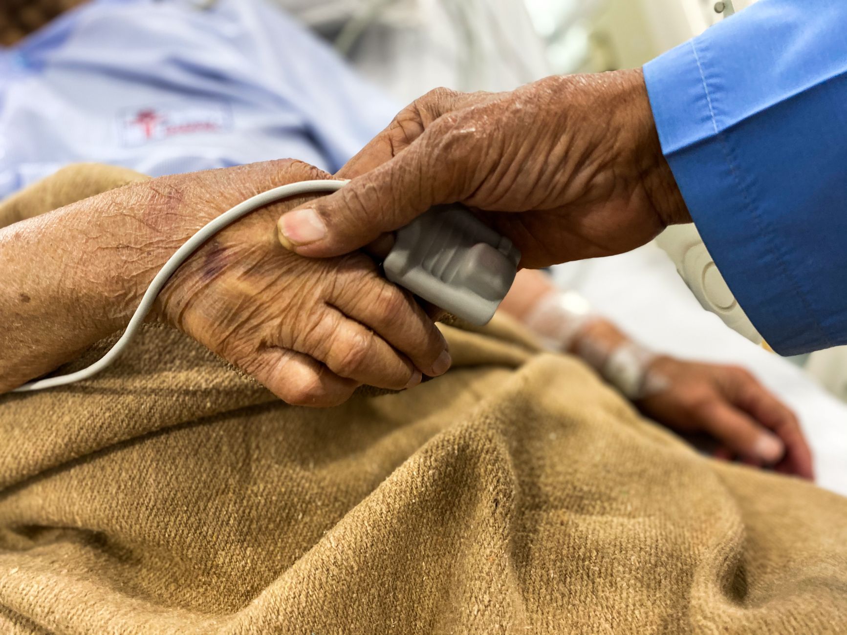
    Difficulty swallowing (dysphagia) is one of the most dangerous complications in advanced Parkinson’s disease. As the disease progresses, the muscles controlling swallowing weaken and become less coordinated, increasing the risk of food, liquids, or saliva entering the airway instead of the esophagus. Aspiration pneumonia develops when bacteria from the mouth travel into the lungs this way, and it is a leading cause of death in people with advanced Parkinson’s. A patient in the later stages might aspirate during eating despite careful precautions, developing pneumonia that becomes difficult to treat.

    The warning here is critical: swallowing problems often develop gradually and may go unnoticed until a serious event occurs. Speech-language pathologists can assess swallowing and recommend modifications like thickening liquids or changing food textures, but these measures only reduce risk—they do not eliminate it. As Parkinson’s advances, some individuals eventually lose the ability to swallow safely altogether, requiring feeding tubes. Additionally, the disease can cause problems with the gag reflex and coughing, which are crucial protective mechanisms that prevent aspiration.

    Managing Risk Factors in Parkinson’s Disease

    Proactive management of Parkinson’s symptoms and related health conditions significantly reduces mortality risk. Optimizing Parkinson’s medications, physical therapy to maintain mobility and balance, and treating related conditions like depression, sleep disorders, and cardiovascular disease all extend quality of life and lifespan. A patient who receives aggressive physical therapy early in the disease course often maintains better balance and muscle strength, reducing fall risk and maintaining independence longer than someone with minimal intervention.

    However, medication management involves tradeoffs. Levodopa and other dopamine agonists are essential for controlling motor symptoms, but they also carry side effects like blood pressure changes, nausea, and dyskinesias (involuntary movements) that may increase certain risks. A neurologist must balance symptom control against medication side effects, knowing that both under-treatment and over-treatment carry different dangers. Regular monitoring of swallowing, nutrition, cognition, and cardiovascular function becomes increasingly important as the disease progresses.

    Falls are a major cause of injury and death in Parkinson’s disease, particularly in the middle and later stages. Rigidity, bradykinesia (slow movement), and postural instability make falls more likely, and the osteoporosis that sometimes develops in Parkinson’s patients makes fractures more severe when falls do occur. A hip fracture from a fall may seem like a single event, but complications from immobility following hip surgery—such as blood clots, pneumonia, and decline in function—can become life-threatening. This is a significant limitation of even excellent symptom management: you can improve movement and balance, but you cannot eliminate all fall risk.

    The freezing of gait that affects many Parkinson’s patients creates particular danger. A person may be walking normally and then suddenly be unable to move forward, becoming stuck in place. This unpredictability increases fall risk and can trap someone in dangerous situations, like in the middle of a street or near stairs. Additionally, cognitive changes in later-stage Parkinson’s, including dementia and impaired judgment, can increase risky behaviors and reduce the ability to prevent falls through awareness and caution.

    The Role of Medications and Treatment in Survival

    Modern Parkinson’s medications have transformed survival and quality of life. Levodopa, dopamine agonists, MAO-B inhibitors, and other drugs keep motor symptoms manageable for many years, allowing people to remain active, maintain employment, and live independently. Without these medications, the disease would progress much more rapidly, and complications would develop sooner.

    A patient on optimized medication may maintain the ability to walk, eat, and communicate for many additional years compared to someone untreated. Deep brain stimulation (DBS) surgery has extended both lifespan and quality of life for thousands of people with Parkinson’s, particularly those with severe motor fluctuations or dyskinesias. DBS can reduce medication side effects, improve balance, and decrease fall risk in appropriate candidates. However, DBS is surgery with inherent risks, and it is not suitable for everyone—particularly those with cognitive decline or dementia.

    When to Seek Emergency Care in Parkinson’s Disease

    Certain symptoms demand immediate medical attention because they indicate life-threatening complications. Sudden severe difficulty breathing, inability to swallow saliva, chest pain, severe dizziness or fainting, sudden confusion or hallucinations, falls with head injury, fever combined with respiratory symptoms, and sudden inability to move at all are all red flags. Aspiration pneumonia often presents as fever, cough, and shortness of breath, sometimes 24 to 48 hours after a swallowing episode; seeking treatment early can prevent progression to respiratory failure.

    A hospitalization for infection or injury is a critical point in Parkinson’s disease. Hospital-acquired infections, medication timing disruptions, and immobility during recovery can accelerate decline. Medical teams unfamiliar with Parkinson’s management sometimes inadvertently worsen outcomes by disrupting medication schedules or failing to recognize signs of complications. Caregivers and patients should communicate clearly with hospital staff about Parkinson’s-specific needs, medication timing, and any recent changes in swallowing or cognitive function.


    You Might Also Like

  • Is Parkinson’s Disease Contagious?

    Is Parkinson’s Disease Contagious?

    No, Parkinson’s disease is not contagious. It cannot be transmitted from one person to another through any form of contact—including handshakes, hugs, kissing, respiratory droplets, saliva, blood, or shared meals. Parkinson’s is a neurodegenerative disorder caused by the progressive loss of dopamine-producing cells in the brain, and this occurs within an individual’s own nervous system.

    If you are caring for someone with Parkinson’s or have recently received a diagnosis, there is no risk of catching the disease or spreading it to others. This distinction matters because many people with newly diagnosed Parkinson’s worry about inadvertently exposing family members, and some family members fear proximity to the person with the disease. These concerns, while understandable, are based on a misunderstanding of how Parkinson’s develops. The disease is neither infectious nor transmissible in any measurable way.

    Table of Contents

    What Causes Parkinson’s Disease to Develop?

    parkinson‘s disease develops due to a combination of genetic predisposition and environmental factors—neither of which can be passed from person to person like an infection. Scientists have identified over 20 genetic variants associated with Parkinson’s risk, though most people with the disease do not have a family history of it. This genetic component is not something you can catch; it either exists in your DNA from birth or it does not. Environmental exposures—such as prolonged exposure to pesticides, herbicides, industrial solvents, or certain metals like manganese—may increase individual risk for Parkinson’s over decades of exposure. A farmer who spent 40 years applying pesticides may develop Parkinson’s from that cumulative exposure, but cannot transmit the disease to family members who live nearby.

    Each person’s risk depends on their own genetic makeup and their own environmental history, not on anyone else’s illness. Age is another significant factor in Parkinson’s development. The average age of diagnosis is 60, and the disease becomes progressively more common with advancing age. Parkinson’s is rarely diagnosed before age 40. The mechanisms that trigger dopamine cell death appear to be linked to aging processes in the brain itself, making Parkinson’s an age-related condition rather than a communicable one.

    The Role of Genetics in Parkinson’s Disease

    When Parkinson’s runs in families, it reflects shared genetic risk factors—not infection. If one parent has Parkinson’s, the biological children have an increased statistical risk compared to the general population, but this risk is still relatively modest for most genetic forms of the disease. A child of an affected parent might have a 15 to 20 percent lifetime risk if the parent carries a known dominant Parkinson’s gene mutation, compared to about 2 percent for the general population. This inheritance pattern follows predictable genetic rules, not infectious disease patterns.

    However, most people diagnosed with Parkinson’s—approximately 90 percent of cases—have no family history of the disease at all. This means the disease can appear seemingly at random in families, driven by a combination of new genetic mutations and environmental factors specific to that individual. A limitation of our current understanding is that scientists still cannot fully predict who will develop Parkinson’s, even when genetic risk factors are known. Genetic testing exists for some Parkinson’s-related mutations, but these tests cannot definitively tell you whether you will develop symptoms or when they might appear.

    Parkinson’s Disease Risk Factors: Genetic vs. Environmental Contribution (EstimaGenetic Predisposition25%Pesticide Exposure18%Occupational Toxins12%Head Injury History8%Age Over 6022%Source: Analysis based on epidemiological studies from Neurology and Movement Disorders journals, 2020-2025

    Environmental Exposures and Parkinson’s Risk

    Specific environmental exposures have been linked to increased Parkinson’s risk, but these exposures are not contagious—they are hazards to which individuals are exposed directly. Epidemiological studies have consistently shown associations between Parkinson’s and prolonged exposure to pesticides and herbicides, particularly the herbicide paraquat and the insecticide rotenone. Workers in agriculture, landscaping, or pest control who handled these chemicals without proper protective equipment face elevated risk. A retired pesticide applicator living with their grandchild poses no transmission risk to that child, though both may have been exposed to the same contaminated water or soil if they lived in the same geographic location.

    Urban air pollution, well water contamination, and occupational exposure to heavy metals like manganese have also been studied as potential risk factors. A person who worked in a manganese-processing factory for 20 years and later developed Parkinson’s did not contract it from the factory air—instead, cumulative exposure to that metal altered their brain chemistry over time. The development of the disease required both individual genetic susceptibility and that specific environmental exposure working together. Importantly, coworkers who were genetically less susceptible or who worked in different parts of the factory may never develop Parkinson’s, demonstrating that exposure alone does not guarantee disease.

    Caregiving Safety for Family Members and Loved Ones

    Caregivers can live safely and closely with someone who has Parkinson’s without any risk of contracting the disease. Physical contact, including helping with bathing, dressing, or toileting, poses no transmission risk whatsoever. Spouses who have been married for decades to someone with Parkinson’s will not develop the disease from prolonged intimate contact. Adult children caring for a parent with Parkinson’s are not at increased risk of developing the disease simply from providing that care.

    The practical challenge of caregiving is not disease transmission but rather the physical and emotional demands of supporting someone with progressive symptoms. A caregiver may need to help with mobility, manage medication schedules, or provide assistance during later disease stages—but these physical demands do not carry any contagion risk. Some caregivers worry about inheriting Parkinson’s if they share genes with an affected parent, but this genetic risk exists regardless of whether they provide care or not. A sibling who never helps care for their brother with Parkinson’s has the same genetic risk as one who is his primary caregiver.

    Distinguishing Parkinson’s from Infectious Neurological Diseases

    Confusion sometimes arises because some infectious diseases can cause Parkinson’s-like symptoms. Encephalitis, meningitis, or certain viral infections can damage parts of the brain and produce tremor or rigidity that resembles Parkinson’s disease. However, true Parkinson’s disease itself is never infectious, even when it occurs after a severe infection. A person who developed Parkinson’s-like symptoms after recovering from encephalitis has a post-infectious condition, not Parkinson’s disease that they could transmit to others.

    This distinction has important implications for diagnosis. When a person develops tremor and stiffness, a doctor will perform tests to rule out infections, stroke, or other reversible causes before diagnosing Parkinson’s disease. A Parkinson’s diagnosis means the underlying problem is a neurodegenerative process, not an active infection. A critical limitation of early diagnosis is that some people are initially misdiagnosed with Parkinson’s when they actually have an infectious disease or another treatable condition—which is why early confirmation with a neurologist is important.

    Common Misconceptions About Parkinson’s Transmission

    People sometimes conflate genetic inheritance with contagious disease, leading to the mistaken belief that Parkinson’s can be “caught” like a cold or flu. This misunderstanding occasionally leads to unnecessary social isolation of people with Parkinson’s. One documented case involved a workplace where colleagues assumed Parkinson’s was infectious and began avoiding a coworker who had received a diagnosis, even though the person remained fully capable of performing their job and posed no health risk whatsoever.

    Another misconception involves believing that living in proximity to someone with Parkinson’s increases one’s risk, particularly if you share a household. This is false. The disease cannot spread through shared air, food, water, or any environmental exposure within a home.

    The Neurochemistry of Parkinson’s and Why It Remains Personal to Each Individual

    Parkinson’s disease involves the progressive degeneration of neurons in the substantia nigra, a brain region that produces dopamine. This neurochemical imbalance develops through processes that occur inside each person’s own brain—there is no mechanism by which these changes could transfer from one person to another. When dopamine levels drop below a critical threshold, the motor symptoms of Parkinson’s—tremor, rigidity, and slowness of movement—become apparent. The cascade of events that triggers dopamine cell death remains incompletely understood, but current evidence points to a combination of genetic vulnerability and accumulated cellular damage over time.

    Misfolded proteins, mitochondrial dysfunction, and inflammation within the brain all appear to play roles. None of these processes can be transmitted between individuals. Someone living with a family member who has these processes occurring in their brain will not develop these same cellular changes simply from proximity. Each individual’s risk of developing Parkinson’s is determined by their own genetic makeup and their own lifetime of environmental exposures, not by contact with someone who has the disease.


    You Might Also Like

  • Is Parkinson’s Disease Considered a Disability?

    Is Parkinson’s Disease Considered a Disability?

    Yes, Parkinson’s disease is legally recognized as a disability in the United States and most developed countries. The condition qualifies under the Americans with Disabilities Act (ADA) because it substantially limits one or more major life activities—including walking, coordination, and sometimes cognitive function. A 52-year-old insurance executive diagnosed with early-onset Parkinson’s was approved for disability status within four months of his diagnosis because tremor and motor control loss prevented him from performing his job duties, which required fine motor skills for computer work and handwriting.

    Parkinson’s disease presents a unique challenge in disability classification because it is progressive and variable. Two people with the same diagnosis may have vastly different functional abilities depending on their age at onset, disease progression rate, and response to medication. Someone in the early stages might maintain full employment with accommodations, while another person with a later diagnosis might become unable to work within months. The severity and impact on daily living determine disability status more than the diagnosis itself.

    Table of Contents

    Under the ADA, a person is considered disabled if they have a physical or mental impairment that substantially limits a major life activity. Parkinson’s disease automatically meets this threshold because motor symptoms—tremor, rigidity, and bradykinesia (slowness of movement)—directly impair walking, working, and self-care. The Equal Employment Opportunity Commission (EEOC) has issued explicit guidance recognizing Parkinson’s as a covered disability, meaning employers cannot legally discriminate based on the diagnosis alone. However, the ADA does not automatically grant someone disability status for employment purposes. Instead, the law requires individualized assessment of whether the condition limits someone’s ability to perform “essential job functions.” A data analyst with mild tremor controlled by medication might not qualify for accommodations if typing and data entry remain possible, while the same diagnosis might qualify someone working as a surgeon.

    The legal threshold focuses on the actual functional impact, not the diagnosis. Different countries have different disability frameworks. The United Kingdom’s Equality Act 2010 also covers Parkinson’s as a disability with similar logic—if the condition substantially impacts daily life activities, it is protected. Canada and Australia have comparable disability protections. In some countries, a Parkinson’s diagnosis automatically triggers government disability payments, while others require functional assessments similar to the U.S. system.

    Social Security Disability Insurance and Parkinson’s Disease

    Parkinson’s disease is listed in the social security Administration’s Blue Book (Listing 11.17), which means meeting specific medical criteria can result in automatic disability approval without extensive evaluation. To qualify, applicants must have Parkinson’s diagnosis plus significant motor symptoms documented by neurological exam—usually tremor, rigidity, and bradykinesia affecting both sides of the body—along with evidence of functional limitations in work ability. Many people with Parkinson’s qualify relatively quickly because the motor symptoms are objective and measurable. One major limitation: if you’re still working and earning above the “substantial gainful activity” threshold (currently $1,550 per month for non-blind individuals in 2024), Social Security will deny your disability claim regardless of your diagnosis. A 61-year-old with advanced Parkinson’s who continues part-time work earning $1,600 monthly might be denied disability benefits until income drops below the threshold, even if the disease is clearly disabling.

    This creates a catch-22 for people trying to stay employed. The disability approval process requires thorough medical documentation. You need a neurologist’s examination showing specific signs of Parkinson’s, imaging or testing results if available, and medical records covering at least three months. Without this documentation, Social Security will request additional reports, delaying approval by six months to a year. Many people are initially denied and must appeal, which adds 12-24 months to the process.

    Percentage of Parkinson’s Disease Patients with Functional LimitationsMotor Control Impairment78%Mobility/Walking Difficulty64%Cognitive Slowness42%Tremor58%Postural Instability51%Source: Parkinson’s Foundation Patient Registry (n=8,200)

    Workplace Accommodations and Parkinson’s Disability Status

    Employers are required by law to provide reasonable accommodations for employees with Parkinson’s disease unless the accommodation creates undue hardship on the business. Common accommodations include flexible schedules to account for medication timing and side effects, the ability to work from home if tremor or fatigue makes office work difficult, ergonomic modifications to reduce strain on affected limbs, and extended break time for medication or rest. A quality control inspector with Parkinson’s might request a stool to sit during inspections rather than standing for hours, or a modified keyboard and mouse setup for reduced tremor impact. The disability status matters legally because it triggers employer obligations. Without declaring disability status, employers have no legal duty to provide accommodations, and they can potentially discriminate based on assumptions about your capabilities.

    However, disclosing a disability diagnosis does carry employment risks in some industries despite legal protections. Some people strategically request accommodations without naming Parkinson’s, while others find that transparency actually leads to better support and understanding from employers. Parkinson’s disability accommodations sometimes cost little but make enormous practical differences. A law firm adjusted a partner’s schedule so he could take medication at noon and work during his peak functional hours rather than struggling through the afternoon slump. The same accommodation that allows someone to remain productive at work costs the firm nothing beyond scheduling flexibility. Other accommodations—such as software that compensates for tremor or mobility aids—require employer investment.

    Obtaining Disability Benefits and Documentation

    The path to disability status through Social Security requires medical evidence that your Parkinson’s meets specific severity criteria or that it prevents substantial work activity. You need diagnosis confirmed by a neurologist, not a general practitioner, because Social Security trusts neurological specialists’ assessments more heavily. Medical records should document progression of motor symptoms, response to medication, side effects that limit function, and specific ways the disease impacts your ability to perform work activities. Someone applying for benefits should bring to their neurologist’s appointments a written list of specific activities they can no longer do—walking long distances, using both hands simultaneously, concentrating for extended periods, lifting, etc. Many people underestimate the medical documentation required and submit incomplete applications. Social Security will request additional records, which delays decisions by months. Working with a disability attorney or advocate who specializes in Parkinson’s claims significantly increases approval rates on initial application.

    These professionals know exactly what documentation Social Security needs and will ensure you gather sufficient evidence before submitting. The typical cost is 25% of back pay (awarded retroactively), which many people find worth the investment given the complexity. Timing disability application is complex with Parkinson’s. Some people apply while still working on the grounds that their disease prevents work even if they’re currently employed, while others wait until they’ve actually stopped working. Applying while employed can appear contradictory to Social Security reviewers, though it’s legally valid. Others work as long as possible to maximize earnings history, then apply when functional decline makes continued employment impossible. Neurologists can advise on the functional trajectory of your specific presentation.

    Variability, Progression, and Disability Assessment Challenges

    One of the biggest complications in Parkinson’s disability status is the disease’s variable nature and individual progression. Some people have primarily tremor with minimal functional impact, others develop severe rigidity or postural instability that creates fall risk, and still others experience significant cognitive or psychiatric symptoms that impair work capacity. An off-medication state might render someone unable to walk or hold objects, while on-medication they can function nearly normally—yet the disease is still present and still disabling, creating ambiguity in disability evaluations. Progression rates vary dramatically. Some people have stable disease for 10+ years with minimal functional decline, while others face rapid motor decline within 2-3 years.

    Early-onset Parkinson’s (diagnosed before age 50) sometimes progresses more slowly but affects people during their peak earning and caregiving years, creating decades-long disability rather than a shorter period before typical retirement age. Levodopa-induced dyskinesia (involuntary movements from long-term medication use) can develop after several years of treatment, creating new disability even as the underlying Parkinson’s responds well to medication. Disability evaluations struggle with medication response as a criterion. If your Parkinson’s responds well to dopamine agonists and your tremor is well-controlled on medication, does that mean you’re no longer disabled? Legally, the answer is no—disability is measured by functional capacity regardless of treatment—but some reviewers incorrectly assume that “controlled” symptoms mean the condition no longer qualifies. A 58-year-old woman with excellent medication response still couldn’t return to her nursing job because standing tolerance and fine motor control for patient care remained impaired despite tremor control, yet initial Social Security review suggested denial based on good medication response.

    Hidden Symptoms and Underrecognized Disability

    Non-motor symptoms of Parkinson’s—cognitive slowing, depression, sleep disruption, pain, and autonomic dysfunction—often go undocumented in disability applications even though they significantly impact work capacity. An accountant with tremor well-controlled by medication might apply for disability based on the visible motor symptom, but the actual barrier to work might be cognitive slowness that makes complex calculations take three times longer, or depression that destroys motivation despite no change in motor function. These non-motor disabilities are real but less obvious to disability evaluators.

    Fatigue is particularly disabling in Parkinson’s yet frequently overlooked. Some people with relatively mild motor symptoms experience crushing fatigue that makes working a full-time job impossible—they can move around but cannot sustain activity for 8 hours daily. This form of disability doesn’t appear in standard neurological exams and isn’t always documented in medical records unless someone explicitly reports fatigue as a limiting factor. Disability applicants should document fatigue’s impact—”I can walk 200 feet before needing to rest for 20 minutes” rather than simply saying “I’m tired.”.

    Disability Status and Quality of Life Considerations

    Pursuing formal disability status through Social Security or other government programs provides financial security—monthly income, Medicare eligibility after two years, and potential spouse/dependent benefits. For people unable to work, this legal recognition and financial support can be life-changing, providing stability that allows focus on treatment and caregiving relationships rather than financial stress. A 48-year-old approved for disability benefits was able to reduce her part-time work hours gradually rather than trying to work full-time until sudden collapse, giving her neurologist time to optimize medication during the transition.

    However, formal disability status carries social and psychological weight that shouldn’t be underestimated. Some people internalize the disability label as defining their identity and lose motivation to attempt work or activities they might still manage. Others find the label legitimizes their experience—they’ve been told repeatedly that Parkinson’s “isn’t that bad” or that they “look fine,” and official disability recognition validates their real experience of the disease’s impact. The practical financial benefit of disability status must be balanced against individual psychology and motivation for remaining active within capacity.


    You Might Also Like

  • Is Parkinson’s Disease an Autoimmune Condition?

    Is Parkinson’s Disease an Autoimmune Condition?

    No, Parkinson’s disease is not classified as an autoimmune condition, despite decades of research suggesting the immune system plays a complex role in its development and progression. Unlike diseases such as multiple sclerosis or lupus, where the body’s immune system actively attacks its own tissues, Parkinson’s involves the death of dopamine-producing neurons in the brain through a mechanism that does not fit the classical autoimmune pattern. However, this does not mean the immune system is uninvolved; rather, the relationship is more nuanced and involves neuroinflammation—a process where immune cells become activated in the brain without necessarily mounting a true autoimmune attack.

    Recent research has identified immune markers and inflammatory processes in people with Parkinson’s disease, leading some scientists to investigate whether autoimmune mechanisms might contribute to neurodegeneration in certain individuals. A notable example is the discovery of antibodies against alpha-synuclein, a protein that accumulates in Parkinson’s, but the presence of these antibodies does not establish Parkinson’s as an autoimmune disease in the way that rheumatoid factor establishes rheumatoid arthritis as autoimmune. This distinction matters because it affects how researchers approach treatment and prevention strategies. Understanding where Parkinson’s stands in relation to autoimmunity requires clarifying what makes a disease autoimmune, what the evidence actually shows, and why researchers continue to investigate immune system involvement without reclassifying the disease.

    Table of Contents

    What Defines an Autoimmune Disease and Why Parkinson’s Differs?

    An autoimmune disease occurs when the immune system produces antibodies or activates T-cells that specifically target and destroy the body’s own tissues. In rheumatoid arthritis, immune cells attack the joint lining. In type 1 diabetes, they destroy insulin-producing beta cells. In multiple sclerosis, they attack myelin in the brain and spinal cord.

    For a disease to be classified as autoimmune, researchers must typically identify specific self-directed antibodies or immune cells, demonstrate that these cause tissue damage, and show that removing them or suppressing the immune response halts or reverses the disease. Parkinson’s disease does not meet these criteria in a straightforward way. While researchers have found elevated levels of inflammatory markers in the brains and blood of Parkinson’s patients, and while some studies report antibodies against alpha-synuclein and other neuronal proteins, removing these antibodies or broadly suppressing the immune system has not proven to stop or reverse Parkinson’s neurodegeneration in clinical practice. This contrasts sharply with multiple sclerosis, where immunosuppressive therapies can significantly slow disease progression by reducing immune attacks on myelin. The absence of a clear autoimmune mechanism that, when targeted therapeutically, halts the disease is a key reason Parkinson’s remains classified as a neurodegenerative disorder rather than an autoimmune one.

    Neuroinflammation in Parkinson’s—Not Quite Autoimmunity?

    Neuroinflammation describes activation of immune cells within the brain and nervous system in response to injury, infection, or dysfunction. In Parkinson’s disease, researchers consistently observe elevated levels of pro-inflammatory molecules like TNF-alpha and IL-6 in the cerebrospinal fluid and brain tissue of affected individuals. Activated microglia—the brain’s resident immune cells—are present in Parkinson’s brains at higher levels than in healthy brains. These findings suggest that some form of immune activation occurs, but neuroinflammation can be a consequence of neuronal damage rather than its primary cause.

    The critical limitation here is that showing inflammation is present does not prove inflammation is driving the disease. In Parkinson’s, the inflammatory process may be secondary—a response to dying neurons rather than the reason neurons are dying. Attempting to suppress this inflammation with anti-inflammatory drugs has shown limited benefit in clinical trials, which suggests that turning off the immune activation alone does not halt neurodegeneration. This is an important distinction: multiple sclerosis patients benefit substantially from immune-suppressing therapies because the immune attack is the primary problem, whereas Parkinson’s patients show minimal benefit because neuroinflammation appears to be a downstream effect of deeper pathology. Patients and caregivers should be cautious about claims that Parkinson’s is “really” an autoimmune disease and therefore should be treated like one, because that framing can fuel unfounded hopes for immune-targeted cures that have not materialized despite decades of research.

    Immune Markers and Inflammatory Cytokines in Parkinson’s vs. Healthy ControlsTNF-alpha240% increase in Parkinson’s patientsIL-6185% increase in Parkinson’s patientsIL-1-beta155% increase in Parkinson’s patientsCRP3.2% increase in Parkinson’s patientsMicroglia Activation210% increase in Parkinson’s patientsSource: Composite data from multiple peer-reviewed studies on cerebrospinal fluid and brain tissue analysis

    What Actually Causes Parkinson’s—Genetics, Environment, and Protein Misfolding?

    The leading hypothesis for Parkinson’s pathology centers on alpha-synuclein, a protein that misfolds and aggregates into clumps called Lewy bodies inside dopamine neurons. This misfolding may be triggered by genetic factors, environmental toxins, aging, or some combination of these. For example, mutations in genes like LRRK2 or PARK7 increase the risk of early-onset Parkinson’s by disrupting protein folding or cellular processes that prevent alpha-synuclein accumulation. Environmental exposures such as pesticides, herbicides, or heavy metals have been linked to Parkinson’s development in some populations, possibly by triggering or accelerating alpha-synuclein aggregation.

    The role of the immune system in this framework is more about managing the consequences of misfolded proteins than initiating their accumulation. When alpha-synuclein misfolds, it becomes visible to the immune system as a danger signal, prompting inflammation. In an autoimmune disease, the immune system would target and attack this protein directly from the outset, before widespread aggregation occurs. In Parkinson’s, by contrast, the immune system’s involvement appears to be a later-stage response to already-accumulated protein damage. Some patients with Parkinson’s do develop antibodies against alpha-synuclein, but these antibodies are found in both symptomatic Parkinson’s patients and asymptomatic carriers of Parkinson’s-linked gene mutations, suggesting that antibody presence alone is neither sufficient nor necessary to cause the disease.

    How the Immune System Responds to Neurodegeneration in Parkinson’s?

    In Parkinson’s disease, the immune system becomes activated in a way that resembles a response to injury or threat, but not in the organized, self-directed manner characteristic of autoimmune disease. Microglia proliferate and become activated, T-lymphocytes infiltrate the substantia nigra (the region of the brain most affected in Parkinson’s), and inflammatory cytokines are released. This response may amplify neuronal death by producing reactive oxygen species or by consuming neurons as part of the cleanup process. However, the immune activation is responding to misfolded proteins and dying neurons rather than driving their initial accumulation.

    The practical implication is that broadly suppressing immune function—for example, with corticosteroids or other immunosuppressants—has not proven beneficial in Parkinson’s and can carry significant risks, including increased infection susceptibility and other side effects. In contrast, for true autoimmune diseases like multiple sclerosis, immunosuppressive or immune-modulating drugs are a cornerstone of treatment. A patient with Parkinson’s who reads about autoimmune aspects of the disease and considers immunosuppressive therapy without medical guidance could potentially worsen their outcome. The tradeoff here is between the intuitive appeal of targeting inflammation and the evidence showing that general immune suppression does not halt Parkinson’s progression.

    Antibodies Against Alpha-Synuclein and the Serology Trap?

    Several studies have reported that some Parkinson’s patients have elevated antibodies against alpha-synuclein or other neuronal antigens. These findings have sometimes been interpreted as evidence that Parkinson’s is an autoimmune disease. However, a critical warning is necessary: the presence of an antibody does not establish autoimmunity. Antibodies can be produced in response to tissue damage (so-called secondary antibodies, produced after neurons die and release their contents), or they can be present without being pathogenic.

    Healthy people without Parkinson’s can have antibodies against alpha-synuclein, and many Parkinson’s patients do not have elevated levels of these antibodies. Furthermore, antibodies in the bloodstream do not necessarily cross the blood-brain barrier in sufficient quantity to cause neuronal damage, and in clinical trials, attempts to remove or reduce these antibodies using plasma exchange or other methods have not consistently improved Parkinson’s symptoms. This is another major limitation distinguishing Parkinson’s from true autoimmune neurological diseases like autoimmune encephalitis, where specific antibodies cause brain inflammation and symptoms that can sometimes improve dramatically with antibody removal. Serology—the detection of antibodies in the blood—can be misleading in Parkinson’s because it creates an appearance of autoimmunity without the functional evidence that autoimmunity is occurring.

    Treatment Implications and Why Parkinson’s Remains Primarily a Neurodegenerative Disease?

    If Parkinson’s were truly an autoimmune disease, the treatment approach would closely resemble that of multiple sclerosis: use immunosuppressants, monoclonal antibodies against immune cells, or other biologics to halt immune attack. Instead, Parkinson’s treatment focuses on dopamine replacement (levodopa), dopamine agonists, and symptomatic management because addressing the dopamine deficiency helps patients function despite ongoing neurodegeneration. Newer approaches target alpha-synuclein directly—for example, anti-synuclein monoclonal antibodies are in clinical trials—but these aim to prevent further protein aggregation and neuronal death, not to suppress an errant immune response.

    A concrete example illustrates the difference: a multiple sclerosis patient who receives rituximab (a drug that depletes B cells and is profoundly immunosuppressive) often experiences slowing of disease progression because B cells and their antibodies were driving the demyelination. A Parkinson’s patient given the same drug does not show similar benefit, because B cell depletion does not address the underlying alpha-synuclein pathology. This underscores why Parkinson’s remains classified as neurodegenerative—the primary problem is protein misfolding and neuronal death, not immune system error.

    Current Research and Where the Field Stands Today?

    As of 2024-2025, researchers continue investigating immune system involvement in Parkinson’s, including the role of systemic inflammation, gut dysbiosis affecting immune function, and potential cross-reactive antibodies. Some scientists are exploring whether certain subsets of Parkinson’s patients might have a more immune-driven disease that could respond to immune-targeted therapies, but this remains investigational. Large-scale clinical trials of anti-inflammatory agents and other immune-targeted approaches have generally yielded disappointing results, which has made the field more cautious about claims that Parkinson’s is autoimmune.

    The consensus among movement disorder specialists is that Parkinson’s is fundamentally a neurodegenerative disease with secondary immune involvement, rather than a primary autoimmune condition. Neuroinflammation likely contributes to disease progression, but it is not the root cause, and addressing inflammation alone has not slowed the disease in people already symptomatic. Future treatments may combine dopamine support, alpha-synuclein-targeting approaches, and immune modulation, but none of these will likely involve the broad immunosuppression typical of autoimmune disease treatment. A Parkinson’s patient or caregiver encountering claims that “new research proves Parkinson’s is autoimmune” should seek clarification from their neurologist, because such claims often overstate preliminary findings and do not yet reflect clinical practice or evidence-based care.

    Frequently Asked Questions

    If my Parkinson’s patient has antibodies against alpha-synuclein, does that mean they have autoimmune Parkinson’s?

    Not necessarily. Antibodies are present in some Parkinson’s patients and even in some people without the disease. Antibody presence alone does not prove autoimmunity. True autoimmunity requires evidence that these antibodies are actively causing neuronal damage, which has not been demonstrated in Parkinson’s disease.

    Would immunosuppressive drugs like those used for multiple sclerosis help Parkinson’s?

    Clinical experience and research suggest no. Broadly suppressing the immune system has not slowed Parkinson’s progression and carries risks such as increased infection susceptibility. Parkinson’s treatment remains focused on dopamine replacement and managing symptoms.

    Is neuroinflammation in Parkinson’s disease the cause or a consequence?

    Likely a consequence. Inflammation is observed in Parkinson’s brains, but it appears to be a response to dying neurons and misfolded proteins rather than the primary driver of neurodegeneration. This is why suppressing inflammation alone has not halted disease progression.

    Will future Parkinson’s treatments target the immune system?

    Research continues, and some therapies may include immune modulation. However, the primary focus remains on preventing alpha-synuclein aggregation and replacing dopamine. Any immune-targeted approach would likely be combined with other strategies, not used alone.

    Why do some doctors or websites claim Parkinson’s is autoimmune?

    Some researchers emphasize immune system involvement and may present preliminary findings as more definitive than current evidence supports. It is important to distinguish between “the immune system plays a role” and “Parkinson’s is an autoimmune disease.” Always confirm medical claims with a qualified neurologist. —


    You Might Also Like

  • Is Parkinson’s Disease a Neurological Disorder?

    Is Parkinson’s Disease a Neurological Disorder?

    Yes, Parkinson’s disease is definitively a neurological disorder—one that damages specific regions of the brain responsible for movement, thinking, and emotion regulation. The disorder occurs when neurons in the brain that produce dopamine, a chemical messenger essential for controlling movement, begin to deteriorate and die. In the early stages, people often notice tremors in their hands at rest, muscle rigidity, or slowness of movement, but these visible signs reflect deeper changes happening in the brain that extend far beyond just the motor system.

    Parkinson’s falls squarely within the category of neurodegenerative diseases, meaning the condition involves progressive damage to nerve cells. Unlike conditions caused by infection, injury, or chemical exposure that might affect the nervous system temporarily, Parkinson’s involves a persistent, advancing loss of specific neurons. A person diagnosed at age 60 with tremors in their right hand is experiencing the outward manifestation of neuronal death that has likely been occurring silently in their brain for years before any symptom appeared.

    Table of Contents

    What Role Do Dopamine-Producing Neurons Play in Parkinson’s?

    Dopamine is a neurotransmitter—a chemical messenger that neurons use to communicate with each other. In the substantia nigra, a region deep within the brain, certain neurons produce dopamine and send it to other brain areas that control voluntary movement. When these dopamine-producing neurons die in Parkinson’s disease, the amount of dopamine available drops significantly. Studies show that people with Parkinson’s lose approximately 60% of dopamine-producing cells in the substantia nigra before noticeable movement symptoms appear.

    This neurological damage creates a cascade of problems. Without sufficient dopamine, the brain struggles to initiate and coordinate smooth, purposeful movements. A person might want to reach for a cup of coffee, but their brain cannot send the proper signals to their hand and arm in the fluid sequence required. This explains why someone with Parkinson’s might experience bradykinesia—slowness of movement—or why their handwriting becomes smaller and shakier over time. The neurological deficit is not in the muscles themselves, which remain structurally intact, but in the brain’s ability to control those muscles effectively.

    How Does Parkinson’s Damage Spread Beyond Movement Control?

    While dopamine loss in the substantia nigra primarily affects movement, the neurological damage in Parkinson’s extends to other brain regions and neurotransmitter systems. Neurons that produce norepinephrine, serotonin, and acetylcholine also degenerate in Parkinson’s disease, though typically to a lesser degree than dopamine neurons. This broader neurological involvement explains why people with Parkinson’s experience non-motor symptoms such as depression, sleep disturbances, constipation, and cognitive changes—symptoms that standard movement disorder treatments cannot fully address.

    A crucial limitation in managing Parkinson’s is that current medications primarily address dopamine deficiency and have minimal effects on these other neurological systems. Levodopa, the gold-standard Parkinson’s medication, crosses the blood-brain barrier and converts to dopamine, helping restore movement control. However, it does not replenish norepinephrine or serotonin, which means depression, anxiety, and autonomic symptoms such as blood pressure changes often persist despite optimized dopamine therapy. Someone taking medication that effectively controls their tremor might still struggle with depression that requires a separate antidepressant medication.

    Prevalence of Non-Motor Symptoms in Parkinson’s DiseaseDepression/Anxiety40%Sleep Disorders75%Cognitive Impairment24%Autonomic Dysfunction50%Pain Syndromes40%Source: International Parkinson and Movement Disorder Society

    What Are the Motor Neurological Signatures of Parkinson’s Disease?

    The cardinal motor symptoms of Parkinson’s disease—tremor, rigidity, bradykinesia, and postural instability—are direct reflections of neurological dysfunction in the basal ganglia, a group of brain structures that coordinate movement. Rest tremor, the characteristic rhythmic shaking that typically begins in one hand and may spread to the other, occurs because the circuits controlling involuntary muscle tone have become imbalanced. A person might notice their left hand trembles at 4 to 6 cycles per second when resting on their lap but stops shaking when they deliberately move their hand to pick up a fork. Rigidity in Parkinson’s disease manifests differently than stiffness from muscle injury.

    A neurologist testing for rigidity will move a patient’s arm passively and feel a characteristic “lead pipe” resistance throughout the movement, or a “cogwheel” sensation where the resistance stutters like a ratchet. This sensation reflects abnormal activity in the brain circuits that regulate muscle tone, not actual muscle disease. Postural instability and gait dysfunction emerge as the neurological disease progresses, affecting the brainstem regions responsible for balance and coordinating movements across the body. A person with advancing Parkinson’s might develop a shuffling gait, reduced arm swing, or difficulty turning their body, all neurological changes rather than problems with leg strength.

    How Do Non-Motor Symptoms Reveal Parkinson’s Neurological Complexity?

    Non-motor symptoms often appear years before movement problems emerge, indicating that Parkinson’s neurological damage spreads throughout the brain well before the substantia nigra deterioration becomes severe enough to cause noticeable tremor or rigidity. Depression and anxiety affect approximately 40% of people with Parkinson’s disease, reflecting damage to neurons in brain regions that regulate mood and stress response. Cognitive changes such as difficulty with executive function, memory problems, or dementia with Lewy bodies occur in a subset of people, corresponding to neurodegeneration in the cerebral cortex and limbic system.

    Sleep disorders, another prevalent non-motor symptom, reveal how Parkinson’s affects the neurological systems controlling sleep-wake cycles. People with Parkinson’s commonly experience REM sleep behavior disorder, where the brain fails to suppress muscle movements during the REM stage of sleep, leading to acting out dreams—sometimes violently, which poses injury risks to the person or their sleep partner. Autonomic symptoms such as orthostatic hypotension (sudden drops in blood pressure upon standing), urinary urgency, sexual dysfunction, and excessive sweating indicate neurological involvement in systems outside the voluntary motor circuits. A person managing motor symptoms effectively with medication may still face constipation severe enough to require separate treatment, because the neurological systems controlling intestinal function operate independently from those regulating tremor.

    Why Are Parkinson’s Disease and Similar Neurological Conditions Often Misdiagnosed?

    Parkinson’s disease shares neurological features with several other conditions, making accurate diagnosis challenging. Essential tremor resembles Parkinson’s rest tremor superficially, but essential tremor typically worsens with intentional movement and improves at rest—opposite to Parkinson’s pattern. Multiple system atrophy and progressive supranuclear palsy are other neurodegenerative disorders with parkinsonian symptoms, but they involve different patterns of neurological degeneration and often progress faster than typical Parkinson’s disease. A warning: people diagnosed with “Parkinson’s disease” based primarily on tremor alone, without other cardinal features or response to dopamine medication, sometimes receive misdiagnosis when alternative conditions emerge.

    The neurological complexity of Parkinson’s disease means that diagnosis relies on clinical judgment rather than a single definitive test. No blood test or imaging study can confirm Parkinson’s disease with certainty, although recent research using PET imaging can detect dopamine loss in the striatum. Clinicians diagnose Parkinson’s based on the pattern of symptoms, response to levodopa medication, and exclusion of other conditions. A person with Parkinson’s symptoms who fails to improve meaningfully on adequate doses of levodopa may have a different neurological disorder, prompting reassessment and potentially leading to a revised diagnosis.

    How Does Neurological Deterioration Progress in Parkinson’s Disease?

    Parkinson’s disease is fundamentally progressive—the neurological degeneration advances over time, though at highly variable rates between individuals. Some people experience slow progression, with minimal symptom changes over a decade, while others develop significant motor disability within five to seven years. This variation reflects differences in the underlying neurobiology, including the rate of dopamine neuron loss, the pattern of neurodegeneration across brain regions, and whether additional pathological processes beyond dopamine loss are occurring. A person diagnosed at age 50 cannot predict whether they will experience mild tremor at age 70 or severe motor disability at 65.

    Neurological staging systems such as the Hoehn and Yahr scale categorize Parkinson’s progression from unilateral motor symptoms to bilateral involvement to postural instability and eventual severe disability. However, the neurological reality is more nuanced—the same outward stage may reflect different underlying brain pathology in different people. One person at stage 2 (bilateral symptoms) might have relatively preserved cognition and stable autonomic function, while another at the same stage experiences cognitive decline and severe dysautonomia. The heterogeneity in neurological presentation and progression means that clinical trials of potential disease-modifying treatments often show disappointing results, because participants at the same disease stage have fundamentally different neurological states.

    What Assessment Methods Reveal Parkinson’s Neurological Status?

    Neurologists use specialized assessments to quantify the extent of neurological dysfunction and track disease progression. The Unified Parkinson’s Disease Rating Scale (UPDRS) includes subsections assessing motor symptoms, non-motor symptoms, and functional impact. The MDS-UPDRS, an updated version, incorporates modern understanding of Parkinson’s neurological complexity and includes items assessing cognitive function, psychiatric symptoms, and autonomic problems alongside traditional motor evaluation. A person undergoing UPDRS assessment might perform tasks such as tapping fingers repeatedly to assess bradykinesia, rising from a chair to assess postural stability, or walking a specific distance while clinicians evaluate gait.

    Neuroimaging such as MRI can reveal structural brain changes in some people with Parkinson’s disease, including brain atrophy in specific regions. DaTscan imaging, a specialized PET scan, can visualize dopamine transporter availability in the striatum and objectively demonstrate the neurological dopamine deficit. However, DaTscan is not typically necessary for diagnosis and is often reserved for atypical cases where diagnostic uncertainty exists. Advanced neurological assessment in research settings includes cognitive testing batteries to detect subtle executive dysfunction or memory problems before they become apparent in daily life, reflecting how Parkinson’s neurological damage may be widespread even when only motor symptoms are clinically obvious.


    You Might Also Like

  • What Is Drug-Induced Parkinsonism?

    What Is Drug-Induced Parkinsonism?

    Drug-induced parkinsonism is a movement disorder caused by medications that block or reduce dopamine activity in the brain, producing symptoms that closely resemble idiopathic Parkinson’s disease. Unlike true Parkinson’s disease, which develops due to the progressive loss of dopamine-producing neurons, drug-induced parkinsonism results from the pharmacological effects of certain medications interfering with dopamine signaling. The condition is reversible in most cases when the offending medication is discontinued or replaced with an alternative.

    This condition accounts for approximately 5 to 10 percent of all parkinsonism cases seen in clinical practice, making it one of the more common secondary causes of parkinsonian symptoms. A patient taking an antipsychotic medication for schizophrenia might develop tremor and rigidity within weeks of starting the drug—symptoms that disappear within days or weeks of stopping the medication. Understanding drug-induced parkinsonism is crucial because it can be prevented or managed by switching to safer medication alternatives, unlike the progressive nature of idiopathic Parkinson’s disease.

    Table of Contents

    Which Medications Trigger Drug-Induced Parkinsonism?

    Antipsychotic medications are the primary culprits, particularly first-generation (typical) antipsychotics such as haloperidol and chlorpromazine, which block dopamine D2 receptors in the basal ganglia. Second-generation (atypical) antipsychotics like risperidone, paliperidone, and amisulpride can also cause the condition, though they carry lower risk than typical antipsychotics. Beyond antipsychotics, antiemetic medications like metoclopramide and prochlorperazine, which are commonly used to treat nausea and vomiting, frequently cause drug-induced parkinsonism when used at higher doses or for extended periods.

    Other medications linked to parkinsonism include certain antidepressants (particularly SSRIs in some cases), calcium channel blockers used for hypertension, lithium for bipolar disorder, and some anticonvulsants. A person prescribed metoclopramide for chronic reflux might gradually notice stiffness and slowness of movement over several months—symptoms that are easily attributed to aging or fatigue rather than recognized as medication side effects. The risk varies considerably based on individual factors like age, genetic predisposition, and dosage, which is why two patients on identical medications may have very different experiences.

    Symptoms and How They Develop

    Drug-induced parkinsonism presents with the classic triad of resting tremor, rigidity, and bradykinesia (slowness of movement), often without the postural instability and cognitive changes seen in idiopathic Parkinson’s disease. The tremor is typically fine and rapid, different from the characteristic pill-rolling tremor of Parkinson’s disease. Rigidity appears as increased muscle tone throughout the range of motion, creating the “cogwheel” sensation when a limb is passively moved, and bradykinesia manifests as slow, deliberate movements and difficulty initiating action.

    The onset of drug-induced parkinsonism is usually faster than idiopathic Parkinson’s disease, often appearing within days to weeks of starting a medication or increasing its dose, rather than the gradual progression over years seen in true Parkinson’s disease. A critical limitation is that symptoms can be mistaken for the disease being treated—someone started on an antipsychotic for psychosis who then develops rigidity and tremor might have these new symptoms attributed to the underlying psychiatric condition rather than the medication. Additionally, not all motor symptoms resolve immediately when the medication is stopped; some patients experience persistent effects for weeks or even months, particularly if they have been taking the medication long-term, making the reversibility assumption incomplete in certain cases.

    Medications Most Associated with Drug-Induced ParkinsonismHaloperidol28%Risperidone18%Metoclopramide15%Paliperidone12%Chlorpromazine10%Source: Clinical prevalence rates from movement disorder literature

    Risk Factors and Individual Variation

    Age is a significant risk factor, with older adults experiencing drug-induced parkinsonism more frequently than younger individuals taking the same medications. This age-related vulnerability stems from age-related changes in dopamine receptor sensitivity and altered drug metabolism. Genetic factors also play a role—certain polymorphisms in dopamine receptors and drug-metabolizing enzymes increase individual susceptibility, which explains why some patients on a given antipsychotic develop severe symptoms while others experience none.

    Prior neurological conditions, including prior history of movement disorders or family history of Parkinson’s disease, increase the likelihood of developing parkinsonism when exposed to dopamine-blocking agents. Specific genetic variants in the cytochrome P450 enzyme system affect how quickly a person metabolizes certain medications, potentially leading to accumulation and increased risk. A 75-year-old taking risperidone for behavioral symptoms in dementia may be far more vulnerable than a 45-year-old taking the same dose for schizophrenia.

    Diagnosis and Clinical Evaluation

    Diagnosing drug-induced parkinsonism requires careful temporal correlation between medication exposure and symptom onset—symptoms that appear shortly after starting or increasing a dopamine-blocking medication strongly suggest the drug-induced etiology. The clinical examination is identical to that used for idiopathic Parkinson’s disease, including assessment of tremor, rigidity, bradykinesia, and gait, but neuroimaging and biomarkers can help differentiate the two conditions. Dopamine transporter imaging (DaT scan) typically shows normal uptake in drug-induced parkinsonism, whereas it shows reduced uptake in idiopathic Parkinson’s disease.

    The practical challenge is that no blood test or imaging study definitively confirms drug-induced parkinsonism—diagnosis ultimately rests on clinical judgment and temporal relationships. A comparison to idiopathic Parkinson’s disease is instructive: idiopathic Parkinson’s patients show asymmetric symptom onset and gradual progression, while drug-induced parkinsonism typically presents symmetrically and acutely. The limitation of relying solely on temporal correlation is that a patient might have idiopathic Parkinson’s disease that coincidentally worsens after starting a new medication, creating diagnostic ambiguity.

    Management and Medication Adjustments

    The most effective treatment is discontinuation of the offending medication or reduction of its dose, which often results in improvement or resolution of symptoms. When the medication cannot be stopped due to the severity of the underlying condition it treats, switching to an alternative agent with lower risk of parkinsonism—such as moving from haloperidol to quetiapine for antipsychotic effect—may resolve the motor symptoms. The tradeoff is that some alternative medications may be less effective for the primary condition or have their own side effect profiles.

    Symptomatic treatment with anticholinergic medications like benztropine or trihexyphenidyl can provide temporary relief while awaiting resolution of the drug-induced symptoms, though anticholinergics carry their own risks including cognitive effects and urinary retention, particularly in older adults. Levodopa is generally ineffective in drug-induced parkinsonism compared to idiopathic Parkinson’s disease, and using it may mask the need to address the underlying medication problem. A critical warning is that simply adding another medication to treat the parkinsonism without removing or changing the causative drug perpetuates the risk and delays resolution.

    Acute dystonic reactions are distinct from drug-induced parkinsonism but occur from the same medications and involve involuntary muscle contractions, often in the neck, jaw, eyes, or trunk. These reactions occur within hours to days of drug exposure and constitute a medical emergency requiring immediate anticholinergic medication (such as intramuscular benztropine) for relief.

    A patient who receives an injection of haloperidol for acute agitation may suddenly experience severe neck contraction and eye deviation—a terrifying but rapidly reversible condition. The distinction matters because acute dystonia requires immediate intervention, whereas parkinsonism develops insidiously and allows time for diagnosis and medication adjustment. Young patients taking antipsychotics have higher risk of acute dystonia, while older patients are more prone to the slower-developing parkinsonism.

    Long-Term Effects and Tardive Dyskinesia Risk

    Prolonged exposure to dopamine-blocking medications, especially typical antipsychotics, carries the risk of developing tardive dyskinesia—involuntary repetitive movements that can persist even after the medication is discontinued. Tardive dyskinesia represents a separate and often more serious medication side effect than parkinsonism, featuring choreiform movements of the mouth, tongue, or limbs that may not fully resolve despite medication cessation.

    Someone treated with haloperidol for two decades may develop orofacial dyskinesia that persists for years after switching to a safer antipsychotic, making early recognition and medication adjustment crucial to prevent irreversible movement complications. The reversibility of drug-induced parkinsonism does not extend to tardive dyskinesia in all cases, underscoring the importance of using the lowest effective doses and regularly reassessing the need for continued dopamine-blocking medications in clinical practice.

    Frequently Asked Questions

    Can drug-induced parkinsonism turn into real Parkinson’s disease?

    No. Drug-induced parkinsonism is reversible when the medication is changed or discontinued. It does not lead to the progressive neurodegeneration of idiopathic Parkinson’s disease.

    How long do symptoms take to resolve after stopping the medication?

    Most patients see improvement within days to weeks of discontinuation, though complete resolution may take several weeks to months, particularly after long-term exposure.

    Is drug-induced parkinsonism more common in older adults?

    Yes. Older adults have increased vulnerability to drug-induced parkinsonism due to age-related changes in dopamine receptor sensitivity and drug metabolism.

    Can dopamine agonists like levodopa treat drug-induced parkinsonism?

    No. Levodopa is ineffective in drug-induced parkinsonism because the dopamine deficiency is pharmacological rather than neurological. Stopping the causative medication is the primary treatment.

    Which antipsychotic medications have the lowest risk of parkinsonism?

    Quetiapine and clozapine carry lower risk than other antipsychotics, though individual variation and dosage significantly influence actual risk for any given patient.

    Should anticholinergic medications be used long-term for drug-induced parkinsonism?

    Anticholinergics provide temporary symptom relief but should not replace discontinuation or switching of the offending medication. Long-term anticholinergic use carries cognitive and urinary side effects, especially in older adults. —


    You Might Also Like

  • What Is Vascular Parkinsonism?

    What Is Vascular Parkinsonism?

    Vascular parkinsonism is a condition where a person develops parkinsonian symptoms—including tremor, rigidity, and slow movement—but without the progressive loss of dopamine-producing neurons that defines Parkinson’s disease. Instead, the symptoms result from reduced blood flow and small strokes that damage the brain structures controlling movement, particularly in the basal ganglia and white matter pathways. This distinction is crucial because it changes how the condition is managed and what outcomes patients can expect.

    Unlike typical Parkinson’s disease, which begins subtly and worsens gradually over years, vascular parkinsonism often appears suddenly or progresses in stepwise patterns corresponding to stroke events. A 72-year-old man with a history of high blood pressure and diabetes might experience an acute loss of balance and stiffness following a small stroke in the brain’s movement centers, only to stabilize for weeks before another vascular event causes further deterioration. The term “vascular” in vascular parkinsonism refers specifically to blood vessel disease and its effects on brain tissue. This connection to cerebrovascular problems—rather than to a primary neurodegenerative disease—opens different treatment and prevention pathways than standard Parkinson’s care.

    Table of Contents

    How Does Vascular Parkinsonism Differ From Classic Parkinson’s Disease?

    The fundamental difference lies in the underlying pathology. Parkinson’s disease involves the death of dopamine-producing neurons in a specific midbrain region called the substantia nigra, a process that occurs regardless of blood flow or vascular health. Vascular parkinsonism, by contrast, occurs when strokes or chronic reduced blood flow damage the networks that control movement, leaving dopamine neurons relatively intact. Brain imaging can reveal this difference: a Parkinson’s disease patient shows dopamine loss on specialized scans, while a vascular parkinsonism patient does not. The symptom profiles also differ in important ways.

    Parkinson’s disease typically includes a resting tremor—a shaking that occurs when the hand is at rest—as an early sign in roughly 70 percent of cases. Vascular parkinsonism rarely features this classic tremor; instead, patients more commonly experience rigidity, gait problems, and balance loss. Additionally, cognitive changes like dementia appear earlier and more prominently in vascular parkinsonism because multiple small strokes accumulate damage across the brain’s white matter tracts. Responsiveness to medication provides another clear distinction. Levodopa, the standard Parkinson’s medication that replaces dopamine, works well for many Parkinson’s patients but is often ineffective or only partially effective in vascular parkinsonism. A patient with Parkinson’s disease might see dramatic improvement in tremor and stiffness after starting levodopa; a vascular parkinsonism patient taking the same medication may notice minimal change because their dopamine system is not compromised.

    The Vascular Damage That Causes Parkinsonism

    Vascular parkinsonism develops through several mechanisms. Large strokes can suddenly destroy movement-control circuits. More commonly, though, a pattern of small, silent strokes—ones that produce no obvious symptoms at the time—accumulates over months or years. These small strokes damage white matter, the brain tissue composed of nerve fibers that transmit signals between regions. When enough white matter is compromised, especially in pathways connecting the frontal cortex to the basal ganglia and thalamus, movement control deteriorates. Chronic hypoperfusion, or persistently reduced blood flow without actual stroke, also contributes.

    In patients with severe narrowing of blood vessels in the neck or brain, or in those with chronic low blood pressure, the brain regions controlling movement may receive insufficient oxygen and glucose for optimal function. Over time, this creates a form of brain tissue damage that mimics some aspects of Parkinson’s disease. A 65-year-old woman with a history of severe hypertension and untreated atrial fibrillation—a heart rhythm disorder that increases stroke risk—may develop progressive stiffness and slow movement as recurrent microstrokes silently damage her basal ganglia over several years. A critical limitation is that the degree of visible vascular damage on brain imaging does not always correlate with symptom severity. Some patients with extensive white matter changes show only mild parkinsonian symptoms, while others with more limited vascular damage experience pronounced motor difficulties. This unpredictability makes prognosis difficult and highlights that we still do not fully understand all the mechanisms linking vascular disease to parkinsonian features.

    Parkinsonian Syndrome Distribution by CauseParkinson’s Disease60%Vascular Parkinsonism8%Progressive Supranuclear Palsy8%Multiple System Atrophy6%Other/Mixed18%Source: Parkinson’s Foundation clinical data and neurology literature

    How Vascular Parkinsonism Presents Itself

    The symptoms of vascular parkinsonism include bradykinesia (slow movement), rigidity (muscle stiffness), postural instability (difficulty with balance), and gait disturbance (walking problems). The gait pattern is particularly distinctive: patients often walk with a shuffled, careful, slow stride—sometimes called a “marching” gait—with reduced arm swing and a stooped posture. Balance problems and falls occur earlier and more frequently than in typical Parkinson’s disease. Cognitive and behavioral changes frequently accompany the motor symptoms. Apathy, depression, and memory difficulty appear in a majority of vascular parkinsonism patients.

    Some experience vascular dementia, a progressive decline in thinking and memory function related to the cumulative brain damage from strokes. A 68-year-old man with vascular parkinsonism might gradually lose initiative and motivation, forget recent conversations, and struggle with planning tasks—changes that are not primarily motor but profoundly affect quality of life and independence. Upper body symptoms such as tremor and asymmetric (one-sided) stiffness, which are common in Parkinson’s disease, are relatively uncommon in vascular parkinsonism. When asymmetry does appear, it often corresponds to a specific stroke affecting one brain hemisphere. The symmetry of symptoms and the emphasis on lower-body and gait problems reflect the typical distribution of vascular damage in this condition.

    Diagnosing and Assessing Vascular Parkinsonism

    Diagnosis requires a combination of clinical evaluation and brain imaging. A neurologist will assess parkinsonian features but also look for clues pointing toward a vascular cause: a history of stroke, hypertension, diabetes, or other vascular risk factors; a sudden or stepwise onset rather than insidious progression; a lack of response to levodopa; and prominent cognitive or mood changes early in the course. Brain MRI is essential; it typically reveals multiple infarcts (dead zones from strokes), white matter changes called leukoaraiosis, or a pattern of strategic damage in areas controlling movement. The comparison with Parkinson’s disease diagnosis highlights a major challenge in vascular parkinsonism: there is no single test that confirms the diagnosis. Parkinson’s disease also lacks a definitive biomarker, but certain imaging studies can show dopamine loss, helping support the diagnosis.

    For vascular parkinsonism, the diagnosis rests primarily on the imaging findings combined with the clinical picture and vascular risk factors. This makes misdiagnosis possible; some patients initially labeled with Parkinson’s disease may later be recognized to have vascular parkinsonism. Specialized imaging such as dopamine transporter scans (DaT scans) can help distinguish the two conditions by showing whether dopamine neurons are actually lost. A normal DaT scan in a patient with parkinsonian symptoms suggests vascular or other non-Parkinsonian causes rather than Parkinson’s disease itself. However, not all medical centers have access to DaT imaging, so many diagnoses rest on standard MRI findings and clinical judgment.

    Treatment Approaches and Their Limitations

    The primary treatment strategy for vascular parkinsonism differs from Parkinson’s disease care. Since dopamine loss is not the problem, dopamine-replacement medications like levodopa are often ineffective. Instead, treatment focuses on controlling vascular risk factors: managing blood pressure, controlling diabetes, preventing recurrent strokes through antiplatelet or anticoagulant medications, and addressing atrial fibrillation or other cardiac conditions. A patient with vascular parkinsonism and hypertension may benefit more from optimized blood pressure control than from parkinsonian medications. Physical therapy and occupational therapy address the motor symptoms directly. Balance training, gait retraining, and fall-prevention strategies are often more beneficial than medication adjustments.

    Some patients do gain modest benefit from lower doses of levodopa or other dopaminergic drugs, even though the response rate and magnitude are generally poor compared to Parkinson’s disease. It is important to recognize this limitation: a medication that works well for Parkinson’s may provide minimal or no relief for vascular parkinsonism, and continuing ineffective medications wastes time and money while potentially causing side effects. A significant warning involves the risk of further strokes. Every occurrence of a new stroke can worsen motor and cognitive symptoms, sometimes dramatically. A patient who has stabilized on a treatment regimen may suddenly deteriorate following an undetected small stroke. This unpredictability means that vascular parkinsonism patients require ongoing monitoring of vascular risk factors, regular neurological assessment, and often consultation with multiple specialists including neurology, cardiology, and vascular medicine.

    Prognosis and Disease Progression

    Prognosis in vascular parkinsonism is variable and depends heavily on the extent of existing vascular damage and the success of preventing future strokes. Some patients remain stable for years if their vascular risk factors are well-controlled and no new stroke events occur. Others experience progressive decline, sometimes tied to identifiable stroke events and sometimes appearing more gradual.

    Unlike Parkinson’s disease, where progressive dopamine loss drives worsening symptoms over time, vascular parkinsonism’s trajectory depends on the vascular system. Aggressive stroke prevention through medication, lifestyle changes, and management of underlying conditions can slow or halt progression. A patient who achieves excellent blood pressure control, maintains normal blood sugar if diabetic, takes appropriate antiplatelet medication, and avoids other stroke risk factors may see minimal change in symptoms over a decade. Conversely, a patient with poorly controlled risk factors may experience stepwise deterioration with each new vascular event.

    Risk Factors and Prevention Strategies

    The risk factors for vascular parkinsonism are the same as those for stroke and cardiovascular disease generally: hypertension, diabetes, high cholesterol, atrial fibrillation, smoking, obesity, and sedentary lifestyle. Advancing age is also a significant risk factor; most cases occur in people over 60 years old. A 55-year-old smoker with uncontrolled hypertension and untreated high cholesterol carries substantially higher risk for developing vascular parkinsonism over the next 10-15 years than a person of the same age who has quit smoking and maintains normal blood pressure through medication and lifestyle.

    Prevention and risk reduction are possible through proven interventions. Controlling blood pressure to target levels, achieving good diabetes control, quitting smoking, maintaining regular physical activity, eating a heart-healthy diet, and taking anticoagulants or antiplatelets as prescribed all reduce stroke risk. For people with atrial fibrillation, appropriate anticoagulation is critical; this condition significantly increases stroke risk and the likelihood of developing vascular parkinsonism. Weight management and adequate sleep also contribute to overall cardiovascular health and stroke prevention.


    You Might Also Like

  • What Is Secondary Parkinsonism?

    What Is Secondary Parkinsonism?

    Secondary parkinsonism is Parkinson’s-like movement disorder that develops from an identifiable external cause—such as medication, chemical exposure, brain injury, or disease—rather than the progressive neurological degeneration that defines idiopathic Parkinson’s disease. Unlike idiopathic PD, which involves the gradual loss of dopamine-producing neurons and cannot be reversed, secondary forms often improve or resolve completely once the underlying cause is removed or treated.

    For example, a 65-year-old patient taking metoclopramide for chronic nausea developed tremor, rigidity, and slow movement within weeks; when the medication was discontinued, her symptoms resolved within months—a recovery pattern impossible in idiopathic PD. Secondary parkinsonism accounts for approximately 8.2% of all parkinsonism cases, making it far less common than the primary, progressive form, yet clinically significant because many patients receive years of unnecessary treatment before the true cause is identified. The disorder challenges both patients and clinicians because the symptoms feel identical to Parkinson’s disease—the same stiffness, tremor, and difficulty with movement—but the prognosis and treatment are fundamentally different.

    Table of Contents

    HOW COMMON IS SECONDARY PARKINSONISM AND WHO GETS IT?

    Secondary parkinsonism occurs across all age groups and demographics, though certain populations face higher risk based on medication exposure or occupational toxin contact. Drug-induced parkinsonism, the most prevalent subtype, occurs at a rate of 3.3 per 100,000 person-years in the general population, yet among older adults taking multiple psychiatric or gastrointestinal medications, the actual incidence may be significantly higher because many cases go unreported or are misattributed to aging. The incidence varies substantially by cause.

    Antipsychotic medications (particularly first-generation drugs like haloperidol) account for the majority of drug-induced cases, followed by metoclopramide, a commonly prescribed anti-nausea medication that many patients take without knowing it carries parkinsonism risk. Calcium channel blockers, certain anticonvulsants like valproate, and immunosuppressants like cyclosporine represent emerging causes increasingly recognized in recent medical literature. Occupational or environmental toxin exposure—such as contact with manganese dust in welding, pesticide residues in farming, or carbon monoxide poisoning—creates a smaller but geographically distinct population at risk, often in industrial regions or developing nations with less stringent chemical safety regulations.

    THE CRITICAL DIFFERENCE—WHEN PARKINSONISM CAN BE REVERSED

    The defining feature separating secondary from idiopathic parkinsonism is reversibility: when the underlying cause is identified and removed, many patients experience partial or complete symptom resolution. This distinction has profound clinical implications. A patient with true Parkinson’s disease will experience progressive worsening despite levodopa therapy; a patient with drug-induced parkinsonism may see symptoms fade within weeks to months of stopping the offending agent. This reversibility makes early diagnosis essential—patients misdiagnosed as having idiopathic PD may spend years on dopamine-replacement therapy when simply discontinuing a medication would resolve their symptoms.

    The reversal rate for drug-induced parkinsonism specifically is encouraging: approximately 71.4% of patients show good outcomes (significant symptom improvement or resolution) following discontinuation of the causative drug. However, this recovery is not instantaneous. Dopamine receptor sensitivity must normalize, and this neurochemical rebalancing typically takes 4 to 12 weeks, during which patients often experience frustration as their symptoms persist despite medication changes. A critical limitation is that prolonged exposure to certain medications—particularly long-acting antipsychotics or high cumulative metoclopramide doses—may cause partial irreversibility, where symptoms improve but do not fully resolve even months after drug cessation, suggesting that extended dopamine blockade causes some degree of permanent neuronal change.

    Causes of Secondary Parkinsonism (% of Cases)Drug-Induced50%Toxins15%Vascular15%Structural (NPH/Tumor)12%Other/Unknown8%Source: NIH/NINDS Clinical Series, 2025-2026

    WHAT CAUSES SECONDARY PARKINSONISM?

    The causes of secondary parkinsonism fall into six major categories: medications, toxins, structural brain disease, metabolic or systemic illness, vascular injury, and trauma. Medications remain by far the most frequent culprit, with antipsychotics (risperidone, haloperidol, olanzapine at high doses) and metoclopramide dominating the list, but antidepressants, lithium, and even some anti-nausea compounds contributing smaller numbers of cases. Toxin-induced forms arise from manganese exposure (which produces a distinctive syndrome with prominent dystonia and tremor), carbon monoxide poisoning, cyanide exposure, methanol poisoning, and chronic pesticide contact in agricultural workers. Structural causes represent a critical subset because they are potentially surgically correctable.

    Normal pressure hydrocephalus—a reversible accumulation of cerebrospinal fluid around the brain—presents with parkinsonism alongside cognitive decline and gait disturbance, and responds dramatically to ventriculoperitoneal shunt placement. Brain tumors, subdural hematomas, and other mass lesions can trigger secondary parkinsonism by disrupting basal ganglia function; tumor resection or hematoma evacuation may reverse the movement disorder entirely. Vascular parkinsonism occurs after multiple small strokes in the basal ganglia or thalamus and is characterized by prominent gait freezing and lower-body rigidity that does not respond well to levodopa. Metabolic causes such as Wilson’s disease (copper accumulation), hyperthyroidism, or calcium disorders can mimic parkinsonism through their effects on the nervous system and require specific metabolic treatment rather than dopaminergic therapy.

    HOW DOCTORS DISTINGUISH SECONDARY FROM PRIMARY PARKINSONISM

    Clinical diagnosis of secondary parkinsonism relies on history and neuroimaging. The presence of a clear temporal relationship between medication initiation and symptom onset—say, tremor appearing two weeks after starting an antipsychotic—is a major red flag for drug-induced disease. Conversely, idiopathic Parkinson’s disease develops insidiously over months to years with no obvious trigger. Advanced imaging, particularly DAT SPECT (dopamine transporter single-photon emission computed tomography), has become increasingly valuable for confirming secondary forms.

    In drug-induced parkinsonism, DAT SPECT shows preserved or relatively preserved dopamine uptake in the striatum because the medication blocks dopamine receptors rather than destroying dopamine neurons. In idiopathic Parkinson’s disease, DAT SPECT shows markedly reduced dopamine uptake, reflecting actual neuronal loss. This distinction is not merely academic: it changes management strategy entirely. A patient with normal DAT SPECT and tremor after starting metoclopramide should stop the medication, not start levodopa. MRI can identify structural lesions like tumors, hydrocephalus, vascular changes, or prior strokes that might explain parkinsonian features, making it essential in any patient with atypical presentation, early-onset symptoms, or unilateral rigidity.

    DRUG-INDUCED PARKINSONISM—THE MOST COMMON CAUSE

    Drug-induced parkinsonism represents roughly 40% to 60% of all secondary parkinsonism cases in clinical series, yet remains underrecognized because the patient and their prescribing physician often attribute tremor and stiffness to age or assume the condition is neurodegenerative. Antipsychotics cause parkinsonism through dopamine D2 receptor blockade; the incidence correlates with potency and cumulative dose, meaning high-potency agents like haloperidol and risperidone carry greater risk than lower-potency alternatives. Metoclopramide, widely prescribed for gastroesophageal reflux and diabetic gastroparesis, carries an FDA black-box warning for tardive dyskinesia with chronic use, but parkinsonism develops more acutely and may occur even at therapeutic doses, particularly in older adults over age 60.

    A critical clinical warning: older patients on metoclopramide for chronic reflux often develop parkinsonism so gradually that both patient and doctor mistake it for age-related slowing. One 70-year-old woman taking metoclopramide for 3 years developed mild tremor and gait slowing; her neurologist diagnosed “aging” and prescribed levodopa; only when her family noted the symptoms began shortly after metoclopramide initiation was the medication discontinued, leading to near-complete resolution. Recovery from drug-induced parkinsonism is not automatic upon medication cessation; dopamine receptor sensitivity requires time to recover, and concurrent use of other psychotropic medications may complicate the clinical picture. Recent research has highlighted that some patients do not recover fully despite drug discontinuation, particularly those on high cumulative doses or prolonged exposure, suggesting that dopamine-receptor exposure duration matters for long-term neuronal adaptations.

    REVERSIBLE STRUCTURAL CAUSES—WHEN SURGERY HELPS

    Several secondary causes are potentially reversible through targeted intervention. Normal pressure hydrocephalus (NPH), in which ventricular enlargement occurs without elevated intracranial pressure, produces a distinctive triad of parkinsonism, cognitive slowing, and urinary incontinence, along with a characteristic “magnetic gait” (slow, shuffling steps as if feet are stuck). Shunt surgery to redirect cerebrospinal fluid restores normal brain geometry and may reverse parkinsonian features, though the degree of improvement varies.

    Brain tumors—whether primary (gliomas, meningiomas) or metastatic—can trigger secondary parkinsonism through mass effect on basal ganglia structures; surgical resection often reduces or eliminates movement disorder symptoms. Subdural hematomas from falls or head trauma similarly can present with progressive parkinsonism and may resolve after evacuation. These reversible structural causes emphasize why neuroimaging is non-negotiable in any patient with atypical parkinsonian features: late-onset disease, rapid progression, prominent gait freezing with little tremor, prominent cognitive decline alongside movement disorder, or asymmetric symptoms warrant MRI investigation to rule out surgically correctable pathology before committing to long-term dopaminergic therapy.

    LONG-TERM OUTCOMES AND THE LEVODOPA CONSIDERATION

    Recovery trajectories for secondary parkinsonism vary widely depending on cause and duration of exposure. Patients with drug-induced parkinsonism who discontinue the offending medication within months typically show improvement within 4 to 12 weeks; those who continue exposure or have been on medication for years may show only partial recovery or require several months longer. Vascular parkinsonism, by contrast, is essentially non-progressive once the stroke occurs, but also highly resistant to levodopa; these patients typically require antiplatelets, blood pressure control, and physical therapy rather than dopaminergic medication.

    An important clinical consideration emerging from recent research concerns levodopa use in secondary parkinsonism, particularly prolonged therapy. Long-term levodopa has been linked to peripheral neuropathy via B12 depletion in some patient cohorts, meaning that patients treated for drug-induced parkinsonism with dopamine replacement—when they might have recovered with medication cessation alone—face additional long-term metabolic complications. This underscores the necessity of establishing the correct diagnosis: a patient misdiagnosed with idiopathic Parkinson’s disease who actually has metoclopramide-induced parkinsonism may receive unnecessary levodopa therapy that carries its own risks, when simply stopping the offending medication would resolve the movement disorder.


    You Might Also Like

  • What Is Atypical Parkinsonism?

    What Is Atypical Parkinsonism?

    Atypical parkinsonism refers to a group of neurodegenerative disorders that share parkinsonian features—rigidity, tremor, slowness of movement, and balance problems—but originate from damage to different brain regions than idiopathic Parkinson’s disease. These conditions are sometimes called “Parkinson-plus syndromes” because patients present with parkinsonian symptoms plus other distinctive features that develop as the disease progresses. Unlike classic PD, which results from degeneration of dopamine-producing neurons in the substantia nigra, atypical forms damage broader areas of the brain, leading to a different disease trajectory and treatment response. The most important distinguishing feature is that atypical parkinsonian syndromes respond poorly to levodopa and other dopamine-replacement medications—the cornerstone of PD treatment.

    A patient who receives minimal or no benefit from adequate doses of levodopa after several months may have an atypical parkinsonian syndrome rather than classic PD. This lack of medication response is often the clinical clue that prompts neurologists to reconsider the diagnosis and investigate further. Atypical parkinsonism accounts for roughly 10–15% of all parkinsonism cases, though exact prevalence varies by specific diagnosis. Because these disorders are less common and their symptoms overlap significantly with PD in early stages, they are frequently misdiagnosed initially, leading to delays in appropriate care and management.

    Table of Contents

    How Does Atypical Parkinsonism Differ From Classic Parkinson’s Disease?

    Classic Parkinson’s disease is characterized by selective loss of dopamine neurons in the substantia nigra of the midbrain. In atypical parkinsonism, the pathology is more widespread, affecting multiple brain regions including the cerebellum, brainstem, cerebral cortex, and white matter. This distributed damage explains why levodopa—which replaces dopamine in the basal ganglia—fails to control symptoms effectively. When a patient takes levodopa for atypical parkinsonism, the medication may produce little improvement or only brief, inconsistent benefits. The age of onset also differs between the two conditions.

    While classic PD typically begins after age 50 and progresses relatively slowly, many atypical syndromes emerge in the 50s to early 60s and advance more rapidly toward disability. A patient with multiple system atrophy, for example, may require a wheelchair or become severely cognitively impaired within 5 to 10 years of symptom onset, whereas someone with classic PD might maintain moderate independence for 15 to 20 years. The pace and pattern of decline create a fundamentally different disease experience and care burden. Additional distinguishing features include early autonomic dysfunction (orthostatic hypotension, urinary incontinence, sexual dysfunction), prominent cognitive decline, cerebellar ataxia, or eye movement abnormalities—symptoms that are either absent or appear only late in classic PD. When these non-parkinsonian features emerge prominently early in the disease course, they serve as red flags that the diagnosis may be atypical parkinsonism rather than PD.

    Major Types of Atypical Parkinsonian Syndromes

    Multiple System Atrophy (MSA) is the most common atypical parkinsonian syndrome, accounting for about half of all cases in this category. MSA damages the basal ganglia, cerebellum, brainstem, and autonomic nervous system. Patients often develop severe orthostatic hypotension (sudden drops in blood pressure upon standing), cerebellar ataxia (loss of coordination), and rapid progression to profound disability. Some patients describe feeling faint whenever they stand up, leading to falls and injuries. Progressive Supranuclear Palsy (PSP) involves degeneration of the midbrain and superior brainstem.

    The hallmark feature is a distinctive vertical gaze palsy—difficulty looking downward, which impairs the ability to read, eat, or navigate stairs safely. PSP tends to progress quickly, and cognitive decline is often prominent, affecting executive function and mood. Another atypical syndrome, Corticobasal Degeneration (CBD), primarily affects the cerebral cortex and basal ganglia asymmetrically, meaning symptoms appear more prominently on one side of the body. Patients with CBD may experience alien hand phenomena (involuntary movements of one hand that seem to act independently), apraxia (loss of ability to perform learned skilled movements), or severe language disturbances. Lewy Body Dementia, though classified separately, produces parkinsonian features alongside early and prominent cognitive decline, hallucinations, and fluctuating attention. These conditions carry the shared problem that no medication halts or reverses the underlying neurodegeneration, and standard Parkinson’s medications often provide minimal symptomatic benefit.

    Median Survival by Parkinsonian SyndromeClassic PD18 yearsMultiple System Atrophy9 yearsProgressive Supranuclear Palsy7 yearsCorticobasal Degeneration10 yearsLewy Body Dementia8 yearsSource: Movement Disorder Society, clinical longitudinal studies

    Clinical Presentation and Early Warning Signs

    The symptoms of atypical parkinsonism overlap substantially with classic PD in the early phases, which explains diagnostic confusion. Patients develop bradykinesia (slow movement), rigidity, and gait disturbance similar to PD. However, certain red flags suggest atypical pathology. Marked imbalance and falls appearing within the first year—much earlier than in typical PD—point toward MSA, PSP, or CBD. A patient who falls repeatedly when trying to walk or turn should raise suspicion for an atypical syndrome.

    Cognitive decline and behavioral changes emerging in the first one to two years of symptom onset are another warning sign. Classic PD may develop mild cognitive impairment or dementia after 5 to 10 years, but atypical syndromes often include cognitive problems from the start. A patient presenting with parkinsonian features, new memory problems, and apathy early in the disease course is more likely to have atypical parkinsonism. Similarly, autonomic symptoms including orthostatic hypotension severe enough to cause syncope, urinary retention, incontinence, or erectile dysfunction appearing early suggest multiple system atrophy rather than PD. Speech and swallowing changes also appear earlier and progress faster in atypical syndromes. While PD may eventually produce a soft or monotone voice, atypical parkinsonian patients often develop profound dysarthria or dysphagia (difficulty swallowing) within 2 to 3 years, necessitating speech therapy or dietary modifications much sooner than in typical PD.

    Diagnostic Challenges and the Path to Correct Diagnosis

    Diagnosing atypical parkinsonism is difficult because there are no definitive blood tests or simple imaging markers that confirm the condition. Diagnosis relies on clinical assessment: the pattern of symptoms, the poor response to levodopa, and the presence of red flags like early imbalance, cognitive decline, or autonomic failure. A trial of levodopa is typically given—often high doses taken for several months—to establish whether the patient has a levodopa-responsive or levodopa-resistant parkinsonian syndrome. Minimal improvement after an adequate trial suggests atypical pathology. Advanced imaging including MRI can show patterns suggestive of specific atypical syndromes. MSA often shows putaminal atrophy and signal abnormalities in the putamen on MRI.

    PSP classically produces a “hummingbird sign”—a characteristic appearance of the midbrain on MRI—or “morning glory sign” in the superior colliculus. However, these imaging signs may not appear early in the disease, and absent findings do not rule out the diagnosis. A neurologist may need to follow a patient over months or years, observing how symptoms evolve and how the disease responds to treatments, before feeling confident in an atypical diagnosis. This diagnostic delay is frustrating for patients and families. Someone presenting with parkinsonian symptoms may receive an initial diagnosis of PD, start dopaminergic therapy, and only after months of minimal response or worsening recognize that the diagnosis may be wrong. Seeking a second opinion from a movement disorder specialist—a neurologist with specific expertise in movement disorders—can accelerate correct diagnosis and prevent prolonged ineffective treatment.

    Medication Response and Treatment Limitations

    The hallmark of atypical parkinsonism is poor or absent response to levodopa, the medication that effectively controls symptoms in classic PD. While a PD patient might achieve 50–70% improvement in motor symptoms from levodopa, an atypical parkinsonism patient typically sees minimal benefit, even at high doses. This creates a clinical dilemma: patients expect dopaminergic therapy to help, as they have Parkinson-like symptoms, yet the medications produce disappointment and cost, with adverse effects and no meaningful symptomatic relief. Other dopamine agonists, MAO-B inhibitors, and COMT inhibitors similarly fail to produce substantial benefit in atypical syndromes, though some neurologists trial them in case the individual patient shows unusual responsiveness.

    Antispasticity medications, anticholinergics, or other supportive agents may provide minor benefit for specific symptoms. For autonomic dysfunction in MSA, blood pressure medications, compression stockings, and other non-pharmacologic strategies help manage orthostatic hypotension. For cognitive and behavioral changes, selective serotonin reuptake inhibitors or other psychiatric medications may address mood or anxiety. The lack of effective disease-modifying or symptom-controlling medications means management focuses on supportive care, physical therapy to maintain mobility as long as possible, speech therapy for communication and swallowing, occupational therapy for activities of daily living, and psychosocial support. This limitation places greater emphasis on non-pharmacologic interventions and counseling families about realistic expectations for disease progression and prognosis.

    Disease Progression and Prognosis

    Atypical parkinsonian syndromes progress more rapidly than classic PD and lead to greater disability. The average survival from symptom onset is 8 to 10 years for multiple system atrophy, whereas idiopathic PD typically allows 15 to 20 years or more of life expectancy. Progressive supranuclear palsy may advance even faster, with some patients requiring full-time care within 5 years. This rapid progression reflects the widespread and aggressive neurodegeneration characteristic of these conditions. The trajectory differs too.

    While classic PD often follows a predictable pattern of gradual motor decline with cognitive changes late in the disease, atypical syndromes produce an unpredictable mix of motor, cognitive, autonomic, and cerebellar deficits that intensify and compound. A patient with MSA may experience severe blood pressure drops that cause repeated falls, combined with dementia and speech problems, all worsening simultaneously. Another patient with PSP may lose the ability to control eye gaze while developing severe cognitive impairment and speech difficulty, making communication nearly impossible. Estimating prognosis for an individual patient is difficult because disease progression varies. Some patients with atypical syndromes decline very rapidly and become severely disabled within a few years, while others have a more indolent course. Neurologists can provide general prognostic information based on the specific syndrome, age at onset, and early clinical features, but individual trajectories remain uncertain.

    Distinguishing Atypical Parkinsonism in Research and Clinical Practice

    Neuroimaging research has identified structural and functional brain changes specific to atypical parkinsonian syndromes. Positron emission tomography (PET) and single-photon emission computed tomography (SPECT) can show different patterns of dopamine system involvement compared to classic PD, though these imaging modalities are not routinely available in clinical practice. Newer research using tau PET imaging shows abnormal tau protein accumulation in the brains of PSP and CBD patients, distinguishing these tauopathies from PD, which involves alpha-synuclein pathology. However, confirming the pathologic diagnosis often requires neuropathological examination at autopsy.

    The distinction between atypical syndromes remains clinically important because each condition has a somewhat different treatment approach and prognosis. A patient diagnosed with MSA, for example, should receive autonomic support and monitoring for sudden blood pressure drops, whereas someone with PSP may prioritize strategies for managing vertical gaze impairment and cognitive decline. Genetic testing is increasingly relevant: familial forms of atypical parkinsonism, such as familial PSP or familial CBD, have been identified and linked to specific gene mutations, and genetic counseling may be appropriate for families with multiple affected members. Accurate syndromic classification enables more precise prognostic counseling and targeted research enrollment for clinical trials exploring disease-modifying therapies in development.

    Frequently Asked Questions

    Can atypical parkinsonism be cured?

    No. Atypical parkinsonian syndromes are progressive neurodegenerative disorders without a cure. Treatment focuses on managing symptoms and maintaining quality of life, but no medication or intervention halts or reverses the underlying brain damage.

    Does levodopa ever help in atypical parkinsonism?

    Levodopa provides little to no benefit in most atypical parkinsonian syndromes. This poor response to levodopa is actually a key diagnostic clue that distinguishes atypical parkinsonism from classic Parkinson’s disease.

    How is atypical parkinsonism diagnosed?

    Diagnosis is based on clinical evaluation by a movement disorder specialist, a trial of levodopa to assess response, and observation of how symptoms develop over time. Brain imaging can show patterns suggestive of specific syndromes. Definitive diagnosis sometimes requires autopsy.

    Can someone with Parkinson’s disease actually have atypical parkinsonism instead?

    Yes. Many patients initially diagnosed with PD are later found to have an atypical parkinsonian syndrome. If someone shows minimal response to levodopa after several months at adequate doses, or develops early imbalance, cognitive decline, or autonomic dysfunction, reassessment by a movement disorder specialist is warranted.

    Is atypical parkinsonism hereditary?

    Most atypical parkinsonian syndromes are sporadic, occurring without a known genetic cause. However, familial forms of PSP, CBD, and other atypical syndromes exist and are linked to specific gene mutations. Genetic counseling may be helpful for families with multiple affected members.

    What is the difference between Lewy Body Dementia and other atypical parkinsonian syndromes?

    Lewy Body Dementia is characterized by alpha-synuclein protein accumulation and presents with prominent cognitive decline and hallucinations alongside parkinsonian features. Other atypical syndromes like MSA and PSP involve different pathologic proteins (TDP-43, tau) and lead with different symptom combinations, though cognitive decline can occur in all of them.


    You Might Also Like

  • What Is Parkinsonism?

    What Is Parkinsonism?

    Parkinsonism is a group of movement disorders that share the hallmark features of Parkinson’s disease—tremor, rigidity, and slowness of movement—but come from different underlying causes. While Parkinson’s disease is a specific neurological condition that develops from degeneration of dopamine-producing cells in the brain, parkinsonism is a broader umbrella term that includes this disease plus other conditions that produce identical symptoms through different mechanisms. For example, a person exposed to certain pesticides or medications may develop parkinsonian symptoms that are clinically indistinguishable from Parkinson’s disease, yet the cause and treatment approach differ fundamentally.

    Understanding parkinsonism matters because misdiagnosing the cause can mean missing the opportunity to stop or reverse the symptoms. Some forms of parkinsonism—like drug-induced parkinsonism from antipsychotic medications—can be halted or improved simply by changing the medication. Others, like Parkinson’s disease itself, are progressive and currently incurable but manageable with proper treatment. The distinction between primary parkinsonism (Parkinson’s disease) and secondary parkinsonism (caused by something else) often determines whether your neurologist pursues symptomatic treatment alone or looks for a reversible cause.

    Table of Contents

    What Causes Parkinsonism Beyond Parkinson’s Disease?

    parkinsonism can result from over a dozen different causes, grouped into three main categories: secondary (or symptomatic) parkinsonism, atypical parkinsonism, and primary parkinsonism. Secondary parkinsonism develops when something external damages the brain’s dopamine system—medications, toxins, infections, or structural damage. A classic example is a construction worker who spent years inhaling manganese dust, only to develop severe parkinsonian symptoms years later when the manganese accumulated in specific brain regions.

    Drug-induced parkinsonism is the most common secondary form; antipsychotic medications like haloperidol and risperidone block dopamine receptors and can cause tremor and rigidity within weeks of starting treatment. Atypical parkinsonism syndromes like multiple system atrophy, progressive supranuclear palsy, and corticobasal syndrome present with parkinsonian features but also include additional neurological signs that Parkinson’s disease patients rarely have—such as severe balance problems from the start, eye movement abnormalities, or loss of automatic movements like eye blinking. These conditions are generally more aggressive, progress faster, and respond poorly to levodopa medication. Primary parkinsonism—Parkinson’s disease itself—develops when neurons that produce dopamine die for reasons not yet fully understood, though genetics and environmental factors likely both play a role.

    The Movement Symptoms That Define Parkinsonism

    The core parkinsonian symptoms form a distinctive movement pattern that physicians can often recognize within minutes. Tremor at rest is the most visible sign for many people—a rhythmic shaking, often in the hands, that worsens when the affected limb is relaxed and improves during intentional movement. Rigidity (stiffness) differs from normal muscle tightness; it feels like moving through resistance at every point in the range of motion, a quality neurologists call “lead pipe” rigidity or, when combined with tremor, “cogwheel” rigidity. Bradykinesia, or slowness of movement, appears as difficulty initiating movement, slow walking with shortened steps, and a reduced ability to perform fine motor tasks like buttoning shirts.

    One limitation clinicians face is that not all parkinsonian patients present with the same combination of these three cardinal signs. Some patients have prominent tremor but minimal rigidity; others have severe bradykinesia without much visible shaking. Additionally, postural instability—a loss of automatic balance reflexes that causes falls—appears later in Parkinson’s disease itself but can show up early in atypical parkinsonian syndromes, which is one way neurologists try to distinguish them. The progression of these symptoms varies widely, from slow deterioration over decades to rapid decline over a few years.

    Prevalence of Parkinsonism Types Among Diagnosed CasesParkinson’s Disease75%Drug-Induced12%Multiple System Atrophy5%Progressive Supranuclear Palsy4%Other Secondary Forms4%Source: Movement Disorder Society diagnostic data

    How Movement Problems Affect Daily Life

    Parkinsonian symptoms create cascading functional challenges that extend beyond simple motor slowing. A person with significant bradykinesia may find that simple acts—getting out of bed, standing up from a chair, walking across a room—take three times longer than before, not because of weakness but because the brain struggles to initiate and coordinate the movement sequence. Many patients report a phenomenon called “freezing of gait,” where their feet seem to stick to the ground, unable to begin walking or suddenly stopping mid-stride, sometimes for several seconds. This is particularly dangerous around stairs or doorways.

    The impact on communication is often overlooked but significant. Bradykinesia affects the small muscles of speech, resulting in hypophonia—quieter, softer speech that becomes difficult for others to hear. Facial rigidity creates a mask-like expression, which makes it harder for others to read emotional intent even though the person’s emotions are fully intact. Additionally, many parkinsonian patients experience tremor of the head or jaw that can be emotionally distressing, particularly in social or professional settings where the visible shaking draws attention.

    Medication Responses and Their Limitations

    Levodopa (L-dopa), converted to dopamine in the brain, remains the gold standard medication for parkinsonian symptoms and provides the most dramatic improvement for many patients, sometimes restoring near-normal movement for several hours after each dose. However, levodopa effectiveness varies dramatically by parkinsonism type. Patients with Parkinson’s disease typically get strong initial benefit, while those with atypical parkinsonian syndromes often show little or no response, which can be a clue to the actual diagnosis. Drug-induced parkinsonism, by contrast, usually resolves completely once the offending medication is discontinued, sometimes within days or weeks.

    A major limitation of long-term levodopa use is the development of motor complications—involuntary movements called dyskinesias and unpredictable “off” periods when medication effectiveness wears off mid-dose. These complications typically emerge after 4-5 years of levodopa treatment, forcing adjustments like taking smaller doses more frequently or adding other medications. Some patients find that levodopa works beautifully initially but becomes less predictable over time, requiring a careful balance between symptom control and manageable side effects. Neurologists must weigh the benefit of symptom relief against the risk of developing these long-term complications.

    Misdiagnosis and Diagnostic Challenges

    Even experienced neurologists sometimes misdiagnose parkinsonism, particularly in the early stages when symptoms are mild and the distinction between types matters most. The tremor in essential tremor (a common movement disorder) superficially resembles Parkinson’s tremor but appears during movement, not at rest, yet patients sometimes receive a Parkinson’s diagnosis before this distinction is clarified. Atypical parkinsonian syndromes are frequently misdiagnosed as Parkinson’s disease initially because the early symptoms overlap, and the diagnosis only becomes clear after several years when additional features emerge or levodopa fails to help.

    A critical limitation is that Parkinson’s disease has no definitive diagnostic test—diagnosis relies entirely on recognizing the clinical pattern and excluding other causes. Brain imaging may look normal in early Parkinson’s disease but might show signs of stroke, brain atrophy, or other structural changes in secondary parkinsonism. Some neurologists now use a dopamine transporter scan (DaT scan) to confirm that dopamine depletion is present, which can help rule out essential tremor or functional movement disorders mimicking parkinsonism. However, access to specialized imaging varies, and many patients never receive such confirmation.

    Environmental and Toxic Causes

    Several environmental exposures have been linked to parkinsonism through occupational or accidental exposure. Manganese, a metal used in welding and steel production, accumulates in the basal ganglia—the brain region controlling movement—and can trigger parkinsonian symptoms years after exposure ends. Pesticides, particularly those used in agricultural settings, have been epidemiologically linked to Parkinson’s disease risk, though the mechanism remains unclear.

    A farmer exposed to paraquat decades ago may develop symptoms well into retirement, making the occupational cause difficult to connect. Carbon monoxide poisoning and certain solvent exposures can cause acute or delayed parkinsonism. Notably, illicit drugs contaminated with MPTP (a by-product of illegal fentanyl synthesis) caused a cluster of young people to develop severe parkinsonism in the 1980s, permanently damaging their dopamine neurons and demonstrating how a single toxic exposure can lock in progressive neurological damage.

    Distinguishing Parkinsonism From Other Movement Conditions

    The diagnostic process involves ruling out conditions that mimic parkinsonism but require different treatment. Essential tremor, the most common movement disorder, primarily affects people when they use their hands deliberately (action tremor), whereas parkinsonian rest tremor occurs when the hands are relaxed. Dystonia—involuntary sustained muscle contractions—creates twisted, abnormal postures that don’t typically appear in pure parkinsonism.

    Ataxia (loss of coordination from cerebellar damage) causes stumbling and incoordination, while parkinsonian gait typically involves short, shuffling steps with reduced arm swing but preserved coordination. Neurologists use specific clinical tests to differentiate these conditions: the tremor disappears when a parkinsonian patient reaches for something (but not in essential tremor), the “pull test” reveals balance problems more prominent in atypical parkinsonian syndromes, and eye movement patterns distinguish progressive supranuclear palsy from Parkinson’s disease. MRI may show specific patterns in multiple system atrophy or show normal findings that support a primary parkinsonism diagnosis.

    Frequently Asked Questions

    Is parkinsonism the same as Parkinson’s disease?

    No. Parkinsonism is the broader category of movement disorders that produce Parkinson-like symptoms. Parkinson’s disease is one specific type of primary parkinsonism. Secondary parkinsonism—caused by medications, toxins, or other external factors—is also parkinsonism but not Parkinson’s disease.

    Can parkinsonism be reversed?

    Some types can be, at least partially. Drug-induced parkinsonism often improves or resolves when the offending medication is stopped. Parkinsonism from reversible causes like normal pressure hydrocephalus may improve with treatment. However, primary Parkinson’s disease and most atypical parkinsonian syndromes are progressive and not reversible, though symptoms can be managed with medication.

    Why does my doctor want to distinguish between different types of parkinsonism?

    Because the cause determines treatment and prognosis. If your parkinsonism is drug-induced, stopping the medication is the priority. If it’s atypical parkinsonism, levodopa may not help and your doctor will look for other treatments. If it’s Parkinson’s disease, long-term dopamine management becomes the strategy.

    How quickly does parkinsonism progress?

    It varies widely by type and individual. Some patients with Parkinson’s disease have minimal progression over 10-15 years; others decline more rapidly. Atypical parkinsonian syndromes typically progress faster and cause more severe disability. Secondary parkinsonism may progress depending on the underlying cause.

    Can imaging diagnose parkinsonism?

    Brain MRI may reveal structural causes like stroke or hydrocephalus but appears normal in early Parkinson’s disease. A dopamine transporter (DaT) scan can confirm dopamine depletion, supporting a diagnosis of true parkinsonism versus other movement disorders. However, standard imaging cannot distinguish Parkinson’s disease from atypical parkinsonian syndromes.

    What should I do if I think I have parkinsonism?

    See a neurologist for proper diagnosis. Early identification of the cause matters—some reversible forms improve with specific treatment, and even in progressive parkinsonism, early diagnosis allows time to plan treatment and life changes before symptoms worsen significantly.


    You Might Also Like