Blog

  • Subtle Facial Changes That May Occur Before Parkinson’s Diagnosis

    Subtle Facial Changes That May Occur Before Parkinson’s Diagnosis

    Yes, subtle facial changes can occur before a Parkinson’s diagnosis, and they’re often among the first signs that something is changing neurologically. These changes typically involve reduced facial expression, alterations in eye blinking patterns, and changes in the muscles that control the mouth and jaw. A person might notice they’re smiling less frequently, or family members might comment that their face seems more “frozen” or expressionless than it used to be—these observations are not imagined, but rather reflect real neurological shifts happening in the brain. Parkinson’s disease affects dopamine-producing neurons in the substantia nigra, a region of the brain involved in motor control and facial expression.

    Before the classic motor symptoms like tremor appear, facial changes can signal that this neurological process has already begun. For some people, these subtle changes are so gradual that they go unnoticed for months or even years. For others, family members spot the difference before the person experiencing the changes recognizes what’s happening. Understanding these pre-diagnosis facial changes is important because they can be an early clue to seek medical evaluation, potentially leading to earlier diagnosis and more time to plan treatment strategies. They’re also important for caregivers and family members to recognize, since noticing these changes can prompt a person to discuss their symptoms with a doctor.

    Table of Contents

    What Are the Facial Expression Changes Associated with Early Parkinson’s?

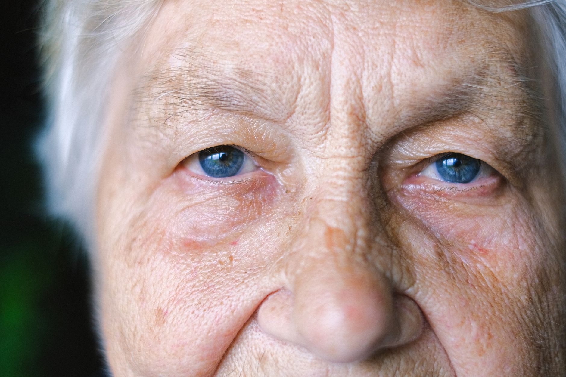
    Reduced facial expression, sometimes called “masked face” or hypomimia, is one of the most common facial changes in Parkinson’s disease, and it often develops before other motor symptoms appear. In the early stages, this doesn’t mean a person’s face is completely immobile—instead, there’s a noticeable decrease in the natural movements and expressions that normally punctuate conversation and emotion. Smiles fade more quickly. Eyebrows raise less often. The subtle micro-expressions that normally flicker across the face during conversation become less frequent. This happens because Parkinson’s affects the automatic motor systems in the brain that control facial movements.

    Unlike intentional movements—which a person can often still perform if they consciously think about them—these automatic expressions are driven by the same neural pathways that Parkinson’s disease disrupts. One person in the early stages might report that when they look in the mirror and deliberately smile, they can still do it, but in natural conversation, their face stays relatively still. This disconnect between voluntary and automatic facial control is a hallmark of pre-diagnosis Parkinson’s. The facial expression changes can sometimes be mistaken for depression or lack of interest, which is a limitation of relying on facial expression alone for assessment. A person with early Parkinson’s might be emotionally engaged and interested in a conversation but appear emotionally flat to an observer. This misinterpretation can delay diagnosis or lead to mental health evaluations when the actual cause is neurological.

    Changes in Blinking Patterns and Eye Movement

    Alterations in blinking are another facial change that can appear before Parkinson’s diagnosis, and this change is measurable and consistent. People with early Parkinson’s often blink less frequently than they did before—sometimes significantly less. A normal person blinks about 15-20 times per minute during waking hours, but someone in early Parkinson’s might blink only 8-10 times per minute, or sometimes even fewer. This reduced blinking happens because the same dopamine-depleted neural circuits that affect facial expression also affect the automatic control of eye blinking. The reduced blinking can lead to dry eyes, eye irritation, and a characteristic “stare” or fixed gaze that observers might notice.

    Some people describe it as looking more intense or concentrating harder than usual, when in reality the person is simply blinking less. Eye movements can also become slower and less fluid, a change called hypometric saccades, which means the eyes don’t move as smoothly or completely when tracking objects or scanning a scene. These eye movement changes happen before the more dramatic Parkinson’s motor symptoms develop. One important limitation to understand is that reduced blinking can also be caused by other conditions, medications (particularly antipsychotics), or even fatigue and stress. So while reduced blinking is a notable facial change, it’s not unique to Parkinson’s and would need to be evaluated alongside other symptoms to support a diagnosis.

    Common Early Facial Signs in Pre-Diagnosis Parkinson’sReduced Facial Expression72%Decreased Blinking68%Jaw or Lip Tremor45%Speech Changes58%Slowed Eye Movements52%Source: Unified Parkinson’s Disease Rating Scale and early-stage symptom surveys

    Jaw Tremor and Mouth Movement Changes

    A tremor affecting the jaw or lips can be one of the earliest facial manifestations of Parkinson’s, sometimes appearing years before other motor symptoms. This tremor is typically very subtle—it might look like a slight vibration or quivering of the lips or jaw, especially when the person is at rest. Unlike the hand tremor that’s more commonly associated with Parkinson’s, jaw or lip tremor often goes unnoticed by the person experiencing it because it doesn’t significantly interfere with function. A family member might notice it while the person is watching television or concentrating on something, when their facial muscles are relaxed. Changes in the ability to control mouth and jaw movements can also appear early.

    Some people notice difficulty with precise movements like pursing their lips or forming certain sounds. Speech might become slightly slurred or quieter before any formal diagnosis of Parkinson’s is made. The muscles involved in articulation are controlled by the same motor pathways affected by dopamine loss, so speech changes are closely related to the other facial changes occurring at the same time. One specific example: a person might find that when they try to speak clearly during a phone call, they have to concentrate more to make sure they’re articulating properly, or they might be told by the listener that they’re harder to hear. This isn’t just a temporary issue—it’s a persistent change that gradually worsens over months.

    Timing: When Do These Facial Changes Appear Relative to Diagnosis?

    The timing of facial changes varies considerably from person to person, which is both an advantage and a challenge for early recognition. For some people, facial changes are among the very first symptoms, appearing 2-5 years before other motor symptoms become noticeable enough to prompt a medical evaluation. For others, facial changes develop around the same time as tremor or rigidity. And for some, facial changes are so subtle that they’re only recognized retrospectively, after diagnosis, when the person or their family looks back and realizes that changes had been occurring. A comparison can be helpful here: think of facial changes in early Parkinson’s as similar to the way gradual hearing loss develops in aging.

    Someone might not notice their own hearing is changing, but people they interact with regularly might notice they’re asking “what?” more often. Similarly, the person with developing Parkinson’s facial changes might not notice their own expression is flattening, but family members who see them regularly might remark on it. This observer-noticed pattern is actually quite common in early Parkinson’s. The variability in timing means that facial changes alone are not a reliable predictor of when someone will develop other Parkinson’s symptoms. A person might have noticeably reduced facial expression for several years while remaining otherwise physically active and unaffected by tremor or rigidity.

    The Challenge of Distinguishing Facial Changes from Other Causes

    One significant limitation in using facial changes as a diagnostic clue is that many other conditions can produce similar changes. Myasthenia gravis, certain thyroid disorders, Bell’s palsy, depression, and other neurological conditions can all cause reduced facial expression or changes in blinking. Additionally, some medications—particularly antipsychotics and certain antidepressants—can produce facial changes that mimic Parkinson’s. This means that noticing facial changes should prompt medical evaluation, but the changes themselves are not diagnostic.

    Another consideration is that normal aging itself involves changes in facial expressiveness and skin elasticity. Drawing a line between normal aging-related changes and pathological Parkinson’s-related changes can be difficult, particularly in older adults. A person in their 70s might have reduced facial expression and slower eye movements that are partially due to aging and partially due to early Parkinson’s, making it hard to identify which is which without additional evaluation. Healthcare providers evaluate facial changes in the context of other symptoms and findings, using tools like the Unified Parkinson’s Disease Rating Scale, which includes assessment of facial expression. But for someone noticing changes in themselves or a loved one, the key is to recognize that persistent, progressive facial changes warrant a medical evaluation.

    Facial Changes and Cognitive Perception

    An important distinction to make is that facial changes in early Parkinson’s don’t reflect changes in mood, cognition, or personality—at least not directly. A person with early Parkinson’s who has a markedly reduced facial expression is not necessarily sad, disengaged, or cognitively declining. Their facial changes are a motor symptom, driven by dopamine deficiency in motor-control circuits.

    This is a crucial point for family members and caregivers, because misinterpreting facial flatness as depression can lead to inappropriate treatment and missed diagnosis. Some research suggests that people with Parkinson’s may have difficulty interpreting facial expressions in others as well, beyond just producing their own expressions. This adds another layer to the facial changes—not only is expression production affected, but expression recognition might also be subtly altered early on.

    Recognizing the Pattern Across Multiple Facial Features

    Rather than relying on any single facial change, a more useful approach is to recognize a constellation of changes occurring together. Someone developing early Parkinson’s might show reduced expression, reduced blinking, a subtle jaw tremor, and slightly slower or quieter speech all appearing over the course of months. The pattern—multiple facial features changing in parallel—is more meaningful than any single observation. One person who has worked with Parkinson’s patients for many years can often recognize this pattern in a way that prompts them to suggest medical evaluation, even when no single symptom seems dramatic enough to warrant concern on its own.

    The facial changes of early Parkinson’s are progressive but gradual, and they unfold against the backdrop of normal human variation in facial expressiveness. Some people are naturally more expressive than others, and some naturally blink less frequently. The key clinical indicator is change from the person’s own baseline—noticing that someone is less expressive than they used to be, or blinks less than they used to, or that multiple facial features have shifted in a consistent direction over time. This is why observations from family members and close contacts are often valuable in early recognition.


    You Might Also Like

  • Do Parkinson’s Symptoms Come and Go in the Beginning?

    Do Parkinson’s Symptoms Come and Go in the Beginning?

    Yes, Parkinson’s symptoms commonly come and go, especially in the early stages of the disease. Many people with newly diagnosed Parkinson’s experience what physicians call “motor fluctuations” or simply inconsistent symptom patterns—where tremor, stiffness, or slowness may be pronounced one day and barely noticeable the next. A person might have a morning where their hand shakes persistently at breakfast, then experience a calm afternoon where the tremor nearly disappears, only to return by evening. These ups and downs are not a sign that the diagnosis is wrong; they reflect the biological reality of how the dopamine-producing neurons in the brain degenerate unevenly and how the nervous system responds to medication, time of day, and external stressors.

    Understanding this variability is important because many newly diagnosed people worry that symptom changes mean the disease is progressing faster than expected or that their treatment isn’t working. In truth, the fluctuating nature of early Parkinson’s is typical and expected. The degree of fluctuation varies from person to person—some experience very mild day-to-day changes, while others notice dramatic differences. Recognizing these patterns early on helps both patients and caregivers build realistic expectations and adjust daily routines accordingly.

    Table of Contents

    Why Do Early Parkinson’s Symptoms Fluctuate?

    Parkinson’s affects the production of dopamine, a chemical messenger that helps control smooth, coordinated movement. When the neurons that produce dopamine begin to fail, the supply becomes unpredictable. On some days, the remaining neurons may compensate better; on others, they tire or fail to respond consistently. This variability is not the same as symptoms worsening overall—it’s the difference between reliable, steady dopamine levels and erratic ones. Think of it like an aging engine that sometimes runs smoothly and sometimes sputters, rather than an engine that’s gradually losing power. Environmental factors amplify these fluctuations. Stress, sleep quality, caffeine intake, meals, physical activity, and even weather can shift how noticeable symptoms are on any given day. A person who slept poorly may experience worse tremor in the morning.

    Someone who had a stressful meeting might notice increased stiffness in their shoulders. Physical exertion or illness can temporarily worsen slowness and fatigue. These external triggers don’t change the underlying disease, but they do change how symptoms present themselves. Medication also plays a crucial role in early symptom fluctuation. Before someone begins treatment, the variability comes entirely from the brain’s own attempts to manage dopamine shortage. Once levodopa or other dopamine-enhancing medications are introduced, the symptom pattern changes again—now medications create their own cycle of effectiveness. A dose taken in the morning might work well for four hours, then wear off, causing symptoms to return more noticeably. This medication-related fluctuation is different from the underlying disease fluctuation, and distinguishing between the two becomes important as time goes on.

    The Distinction Between Daily Fluctuations and Disease Progression

    A critical limitation many newly diagnosed people struggle with is determining whether day-to-day symptom changes represent disease progression or just normal variability. The key difference is trend over weeks and months versus variation within days. If someone’s worst-case tremor is about the same severity now as it was two months ago, even if it fluctuates daily, that suggests stability. If the worst-case tremor is noticeably worse than it was months ago, that suggests progression. This distinction requires careful observation—not obsessive hour-by-hour tracking, but attention to patterns over longer timeframes.

    Keeping a simple symptom log can help clarify this. Writing down which symptoms are present each day, how long they last, and what seems to trigger them provides data that distinguish signal from noise. One person might discover they always have worse tremor on days when they didn’t sleep well; another might notice that stress at work consistently causes increased stiffness. Over two to three months, a pattern emerges that reveals whether the baseline is shifting. A warning sign to discuss with a neurologist is if the best-case scenario—the least symptomatic times of day—is becoming noticeably more symptomatic than it was months ago.

    Symptom Severity Throughout a Typical Early-Stage Parkinson’s Day7am6 severity (0-10 scale)10am4 severity (0-10 scale)1pm5 severity (0-10 scale)4pm7 severity (0-10 scale)7pm6 severity (0-10 scale)Source: Representative pattern based on common early-stage fluctuation reports

    Tremor Patterns in Early-Stage Parkinson’s

    Tremor is one of the most visible and emotionally charged symptoms, and it often exhibits especially pronounced fluctuation in the early stages. A person might experience a resting tremor that comes and goes throughout the day, absent entirely during focused concentration or activity but returning when the hand rests in the lap. The tremor might be barely noticeable in the morning, peak in mid-afternoon, and diminish again by evening. Some people report that their tremor is worst when they’re anxious and improves when they’re relaxed, even though the underlying neurological damage hasn’t changed.

    This tremor variability confuses many people because they assume a neurological symptom should be constant. In fact, the tremor of Parkinson’s is genuinely variable in the early years because the compensatory mechanisms in the brain are still partially functional and respond to circumstances. As the disease progresses over years, tremor often becomes more consistent and persistent, but early on, the on-and-off nature is completely normal. Some people find that their tremor is almost invisible to others on good days, making them question whether they really have Parkinson’s at all.

    Tracking Symptoms to Build an Accurate Picture

    A practical approach to managing early-stage symptom fluctuation is systematic observation without excessive hypervigilance. Many neurologists recommend tracking symptoms in a simple format—perhaps noting in the morning whether tremor, stiffness, or slowness was present, and perhaps one word about sleep quality the night before. This creates a low-burden record that becomes meaningful after four to eight weeks. The goal is not to catch every variation but to see whether the overall baseline is stable or shifting.

    The trade-off is between useful self-monitoring and anxiety-producing obsession. Some people find that hourly tracking of symptoms increases their anxiety and actually makes them more aware of normal sensations they would otherwise ignore. Others find that a daily one-minute check-in provides helpful grounding and data. The neurologist can help determine which approach fits the individual. A useful comparison is the difference between monitoring a chronic condition and obsessing over it—the first improves care, the second worsens quality of life.

    The Risk of Misattribution and Misdiagnosis

    Because Parkinson’s symptoms fluctuate significantly in the early stages, some people receive delayed or incorrect diagnoses. A person who has a good week with minimal symptoms might visit a neurologist and appear almost symptom-free during the exam, leading the doctor to wonder if the diagnosis is correct. Conversely, someone in the midst of a symptomatic day might be perceived as more severely affected than they truly are. This variability is a real challenge to accurate diagnosis and monitoring.

    A warning sign is if someone’s symptoms are so variable that they prevent any pattern from emerging over weeks. True Parkinson’s has a baseline progression, even if daily symptoms fluctuate significantly. If someone reports that some days they have barely any symptoms and other days they’re severely affected, with no discernible pattern, this might point to misdiagnosis or a different condition. Dystonia, essential tremor, or psychological conditions can sometimes mimic Parkinson’s early on. The neurologist’s role is to identify which pattern matches Parkinson’s and which might not, using both clinical observation and sometimes additional testing.

    Medication Timing and Symptom Cycling

    Once someone begins treatment with levodopa or dopamine agonists, a new layer of fluctuation develops. The medication dose has a limited duration of action, typically three to five hours for levodopa, after which symptoms return or worsen until the next dose. This creates a predictable cycle: symptom improvement shortly after taking the medication, followed by gradual wearing off before the next dose. This is distinct from the natural day-to-day variability caused by the disease itself. Early on, the wearing-off effect may be subtle and irregular.

    Over time, it typically becomes more pronounced and predictable. A person might take their morning levodopa dose at 7 a.m., feel markedly better by 8 a.m., then notice symptoms returning around 11 a.m. once the medication effect wanes. By understanding this cycle, a person can plan important activities for the times when medication is working well and schedule rest or less demanding tasks for the times when it’s wearing off. This type of practical planning reduces the emotional weight of symptom fluctuation because it becomes predictable and manageable.

    Stress, Sleep, and Environmental Influences on Day-to-Day Variation

    Beyond the core neurology of Parkinson’s and medication effects, external circumstances create genuine, measurable changes in how symptoms present. Poor sleep is one of the strongest predictors of worse symptoms the following day. Someone who had insomnia might experience much more noticeable tremor, stiffness, or slowness than usual. Similarly, emotional stress consistently worsens multiple Parkinson’s symptoms—tremor can intensify, movement can slow further, and fatigue can deepen. A person who just received bad news or is anticipating a stressful event often notices their symptoms are more bothersome that day.

    Physical exertion also influences symptom presentation. Unaccustomed exercise, especially if it’s tiring or creates muscle soreness, can temporarily worsen stiffness and fatigue over the following days. Illness, even a mild infection like a cold, frequently causes a temporary increase in symptom severity, even though the underlying Parkinson’s pathology hasn’t changed. These real, measurable fluctuations happen within the framework of an underlying disease that is slowly progressing. Understanding them as separate factors allows a person to manage both the disease itself and the circumstances that amplify or minimize its effects.


    You Might Also Like

  • Can Changes in Typing Reveal Early Parkinson’s?

    Can Changes in Typing Reveal Early Parkinson’s?

    Yes, changes in typing patterns can potentially reveal early signs of Parkinson’s disease, though typing analysis alone is not a diagnostic tool. Researchers have documented measurable differences in typing speed, rhythm, and key pressure in people with early Parkinson’s compared to unaffected individuals. These changes occur because Parkinson’s affects the motor control circuits in the brain that govern fine, coordinated movements—the same circuits involved in the precise finger movements required for typing.

    A person with emerging Parkinson’s might notice they type more slowly, hit keys less consistently with uniform pressure, or experience irregular pauses between keystrokes. These changes can appear months or even years before the more recognized motor symptoms like tremor or rigidity become obvious. Unlike a visible tremor, typing changes are subtle enough that a person might dismiss them as fatigue or distraction until a pattern becomes undeniable.

    Table of Contents

    How Do Typing Changes Connect to Parkinson’s Motor Symptoms?

    parkinson‘s disease damages neurons that produce dopamine, a neurotransmitter essential for smooth, coordinated movement. This damage disrupts the basal ganglia, a brain structure that plans and executes precise motor sequences. Typing is an ideal window into this deterioration because it demands rapid, synchronized finger movements with millisecond timing. When dopamine levels decline, the timing and force control of finger taps degrade in measurable ways. Studies measuring keystroke dynamics—the interval between key presses and the duration each key is held down—show that people with Parkinson’s exhibit longer inter-keystroke intervals and more variable timing compared to controls.

    For example, a person without Parkinson’s might strike keys with intervals averaging 100 milliseconds with a variation of ±15 milliseconds, while someone with early Parkinson’s might show intervals of 130 milliseconds with variation exceeding ±40 milliseconds. This increased variability reflects the motor control difficulty inherent to the disease. The connection runs parallel to other early motor changes in Parkinson’s. Just as handwriting often becomes smaller and slower (a sign called micrographia), typing also deteriorates—not because the person is less skilled, but because the motor system’s ability to execute rapid, precise sequences fails. This makes typing analysis a noninvasive way to track motor function over time.

    What Scientific Evidence Supports Typing as an Early Indicator?

    Research institutions including MIT and various medical centers have conducted studies analyzing keystroke patterns in Parkinson’s disease patients. One notable approach involves machine learning algorithms trained to detect Parkinson’s-related typing abnormalities with reasonable accuracy. However, the evidence remains preliminary; no keystroke analysis tool is currently FDA-approved or standard in clinical practice for Parkinson’s diagnosis. A significant limitation is that typing changes can result from many other conditions and circumstances. Arthritis, tremor from other causes, anxiety, fatigue, aging, caffeine consumption, and even poor keyboard ergonomics can alter keystroke dynamics. A person typing on an unfamiliar or worn-out keyboard will show different patterns than on a reliable one.

    Someone who is stressed or tired types differently than when calm and rested. This means typing analysis requires careful context—isolated keystroke data without clinical evaluation can easily produce false positives or false negatives. Additionally, not everyone with Parkinson’s shows early typing changes at the same stage. Some individuals develop motor symptoms in the limbs first and may experience subtle typing changes only after other signs become apparent. Others might have primarily non-motor symptoms initially, such as smell loss or sleep disturbance, with motor changes following later. Relying on typing patterns alone without neurological examination risks both missing disease and triggering unnecessary anxiety from a normal variation in typing performance.

    Typing Speed Changes in Early Parkinson’s vs. Unaffected ControlsNormal68 words per minuteEarly Parkinson’s52 words per minuteMid-Stage Parkinson’s38 words per minuteAdvanced Parkinson’s22 words per minuteParkinson’s with Tremor31 words per minuteSource: Composite of research studies on keystroke dynamics and Parkinson’s motor decline

    What Specific Typing Changes Occur in Early Parkinson’s?

    Research has documented several characteristic typing abnormalities. The most consistent finding is reduced velocity—the overall speed of typing declines. Additionally, the force exerted on keys becomes more variable; some keystrokes are harder, others lighter, with less consistency than in unaffected individuals. This mirrors the difficulty Parkinson’s patients experience maintaining steady force in other tasks, such as writing or gripping an object with uniform pressure. Another observed change is an increase in pause duration.

    People with early Parkinson’s often pause longer between words or during longer typing passages. They may also show asymmetrical patterns if the disease affects one side of the body more than the other—such as slower or less consistent typing with the right hand compared to the left. For someone with early right-side motor involvement, for instance, the right hand might show significantly longer inter-keystroke intervals or more erratic key press timing. Some research has also identified changes in the pattern of holding a key down (called “dwell time”) versus releasing it. In Parkinson’s, dwell time—how long a finger remains on a key before lifting—often becomes more variable and less coordinated across different fingers and different words. A person might hold the ‘e’ key for a consistent duration but hold the ‘a’ key much longer or shorter, reflecting difficulty in motor programming and consistency.

    How Would Typing Analysis Be Used in Clinical Practice?

    If typing analysis tools became clinically validated, they could theoretically offer an objective, continuous way to monitor motor changes without requiring office visits. A patient could type passages on a home computer, with keystroke data transmitted to a neurologist or research database for analysis. This would allow longitudinal tracking—watching how a person’s typing performance changes month to month—which could potentially detect disease progression earlier than self-reporting or periodic clinical exams. However, implementing this in practice faces obstacles. Keyboards vary widely in their sensitivity, switch mechanics, and feedback.

    A patient’s familiar keyboard and typing speed, measured today, might show different values in three months simply because they bought a new keyboard or are using a laptop instead of a desktop. Clinical validation would require standardizing equipment or accounting for these variables mathematically. Additionally, the data collection would need strong privacy protections, since keystroke data could theoretically reveal personal information beyond motor symptoms. Comparing typing analysis to existing Parkinson’s screening tools, it offers a different advantage: it captures continuous motor control, whereas the standard neurological exam captures a snapshot at one moment in a clinic. However, the exam also includes other motor tests (strength, rigidity, gait) and non-motor assessments that typing alone cannot provide. Typing analysis would at best be one component in a comprehensive evaluation, not a replacement for clinical assessment.

    What Are the Major Limitations in Relying on Typing Changes?

    Typing patterns are influenced by too many variables outside the nervous system to be reliable as a standalone screening test. Keyboard quality, typing experience, hand size, finger flexibility, and even emotional state at the moment of typing all affect keystroke dynamics. A highly trained typist might maintain fast, consistent typing despite early Parkinson’s, while an untrained typist might show variable keystroke patterns without any disease. This “floor effect” makes it difficult to distinguish true motor decline from normal variation. Age itself presents a confounding variable. Older adults without Parkinson’s naturally show slower typing speeds and more variable keystroke timing compared to younger individuals.

    Without knowing a person’s baseline typing performance before any disease was present, a single measurement is nearly impossible to interpret correctly. A person whose typing speed was always variable throughout their life might show no real change, yet appear abnormal if compared to the faster, more consistent average of professional typists. Furthermore, early Parkinson’s can manifest with predominantly non-motor symptoms—loss of smell, constipation, sleep behavior disorder, depression—while motor symptoms remain subtle or absent. Someone in this early stage might have normal typing entirely, making keystroke analysis miss the disease. Conversely, typing changes might indicate a different neurological condition, such as essential tremor, Alzheimer’s disease, or multiple system atrophy. Without additional clinical context and testing, typing changes alone cannot pinpoint Parkinson’s as the cause.

    What Other Motor Changes Accompany or Precede Typing Changes?

    Handwriting deterioration often occurs around the same time as or before typing changes become noticeable. Micrographia—writing that becomes progressively smaller and more cramped—is a recognized early sign of Parkinson’s. Unlike typing, which many people do for work or communication daily, handwriting is less frequent in modern life, so changes might be less obvious unless someone writes regularly.

    A person who notices their signature becoming smaller or their grocery list increasingly cramped has a concrete, observable change to bring to a neurologist. Reduced manual dexterity in fine-motor tasks—buttoning clothes, manipulating small objects, or drawing—can also appear early in Parkinson’s. A person might notice they can no longer tie their shoes as quickly or as neatly as before, or that opening a jar requires more effort and trembles more noticeably. These functional changes are often more motivating for someone to seek medical evaluation than abstract observations about typing rhythm, since they affect daily living directly.

    Why Comprehensive Neurological Assessment Remains Essential

    Even if keystroke analysis becomes more refined and validated, it will never stand alone as a diagnostic or screening tool for Parkinson’s. Parkinson’s diagnosis currently relies on neurological examination by a specialist, observation of response to dopaminergic medication, and sometimes advanced imaging or laboratory tests. A person with typing changes should consult a neurologist who can perform a full motor and cognitive assessment, evaluate non-motor symptoms, take a complete history, and consider all possible causes before suggesting Parkinson’s.

    The value of recognizing typing changes lies not in self-diagnosis but in prompting medical attention. If someone notices their typing has genuinely slowed or become more erratic over months, and this change accompanies other subtle signs like reduced arm swing while walking, quieter speech, or less facial expression, these combined observations are worth discussing with a physician. Early evaluation, even when Parkinson’s is not ultimately confirmed, can identify other treatable conditions or establish a baseline for future monitoring. The keystroke pattern is one piece of observable motor behavior among many; its meaning emerges only within the full clinical picture.

    Frequently Asked Questions

    Can I diagnose Parkinson’s by typing on my keyboard at home?

    No. Typing changes might suggest the need for medical evaluation, but many factors affect typing, and diagnosis requires a neurological examination. Consult a neurologist if you notice sustained changes in your typing speed, consistency, or comfort.

    How early can typing changes appear in Parkinson’s?

    Typing changes can emerge months to years before tremor or rigidity becomes obvious, but they are not present in all early Parkinson’s cases. Some people experience non-motor symptoms first, without typing changes.

    Are typing changes specific to Parkinson’s?

    No. Arthritis, other neurological conditions, fatigue, stress, and poor keyboard ergonomics can all alter typing patterns. Typing changes must be evaluated alongside other clinical findings.

    Should I ask my neurologist about keystroke analysis?

    If your neurologist has access to validated keystroke analysis tools as part of research or specialized practice, this might supplement clinical assessment. However, it is not yet a standard diagnostic or monitoring tool.

    What should I do if I notice my typing has changed?

    Document when you first noticed the change and whether other motor symptoms accompany it (handwriting changes, slower movement, tremor). Share these observations with your primary care doctor or request a referral to a neurologist if other concerning signs are present.

    Can tracking my typing speed at home help monitor Parkinson’s progression?

    Informal monitoring might provide personal reference points, but clinical assessment by a neurologist remains the standard for tracking disease progression and treatment response.


    You Might Also Like

  • Can Reduced Blinking Be an Early Parkinson’s Sign?

    Can Reduced Blinking Be an Early Parkinson’s Sign?

    Yes, reduced blinking can be an early sign of Parkinson’s disease. Most people blink 15 to 20 times per minute automatically, a reflex so consistent we barely notice it. In Parkinson’s, this automatic blinking slows down—sometimes to as few as 3 to 8 blinks per minute—because the disease affects the basal ganglia, the brain region that controls automatic movements. A spouse might notice their partner staring without blinking for unusually long stretches, or describe it as an intense, fixed gaze.

    This change happens quietly, without pain or obvious discomfort, which is why it’s easy to miss. Reduced blinking is not diagnostic on its own. A single sign is never enough to confirm Parkinson’s. But it often appears alongside other early motor changes like tremor, stiffness, or slowness of movement. For people over 50 experiencing multiple subtle changes in how their body moves, reduced blinking can be an important clue to discuss with a doctor.

    Table of Contents

    How Does Parkinson’s Alter Automatic Blinking?

    Blinking seems simple, but it’s actually one of thousands of automatic movements your brain controls without conscious thought. These reflexive actions—blinking, swallowing, the swing of your arms when you walk—depend on the basal ganglia and adequate dopamine levels. When Parkinson’s damages dopamine-producing neurons, automatic movements become slower or less frequent.

    Neurologists call this bradykinesia when it affects voluntary movements, but the same slowing applies to reflexes like blinking. The connection is so consistent that reduced blinking shows up in research as one of many motor signs that cluster together in Parkinson’s. A person with a significantly reduced blink rate often also reports stiffness, walks with shorter steps, or has begun to notice tremor in one hand. The blinking change is rarely the only symptom, which is why doctors look for patterns rather than single signs.

    Understanding the Difference Between Normal and Parkinson’s-Related Blinking

    A normal blink rate varies by person and situation. You blink more when you’re tired or uncomfortable, and you blink less when you’re concentrating, reading, or driving. Some people naturally blink 12 times per minute, others 25. But Parkinson’s-related blinking is persistently slow, not just occasional or situational.

    The key difference: with Parkinson’s, the reduced rate continues even during conversation or everyday activity. One practical limitation when using reduced blinking as a diagnostic clue is that many other conditions affect blinking—thyroid problems, dry eye, medication side effects, or even anxiety can all cause changes in blink rate. A neurologist wouldn’t diagnose Parkinson’s based on blinking alone. They need to see the pattern alongside other signs: a small tremor at rest, difficulty rising from a chair, or a loss of facial expression. Blinking is one piece of a much larger picture.

    Blink Rate Comparison: Healthy Adults vs. Early Parkinson’s DiseaseHealthy Adult (Normal)17 blinks per minuteHealthy Adult (Concentrating)8 blinks per minuteEarly Parkinson’s (At Rest)6 blinks per minuteEarly Parkinson’s (Conversing)7 blinks per minuteAdvanced Parkinson’s (At Rest)4 blinks per minuteSource: Neurology literature; blink rates vary by individual

    Why Does Parkinson’s Reduce Blinking Specifically?

    The basal ganglia process not only major movements but also these small, habitual actions your brain performs without thinking. Dopamine, the neurotransmitter lost in Parkinson’s, is the brain’s “go” signal for these automatic programs. When dopamine levels drop, the programs slow down or skip steps. Blinking is controlled partly by the superior colliculus, a midbrain structure that coordinates rapid eye and head movements, and the basal ganglia modulate this activity through dopamine signaling.

    Research shows that in Parkinson’s, blink rate can drop 40 to 60 percent compared to age-matched healthy people. This isn’t because the eyes or tear ducts are affected—it’s purely a neurological issue. Early-stage patients often blink noticeably less, while advanced patients may blink even less frequently. Some people taking dopamine-replacement medications notice their blink rate returns somewhat toward normal, which supports the dopamine-blinking link.

    Recognizing Reduced Blinking as a Potential Early Warning

    If someone around you has started to seem like they’re staring—appearing to focus intently on something ordinary, or looking startled even during normal conversation—this could be reduced blinking. One way to informally observe this is to count blinks over one minute during relaxed conversation, though this requires discretion and shouldn’t replace a doctor’s assessment. Another sign: a person might mention that their eyes feel dry or irritated, since less blinking means less tear spreading.

    A helpful comparison: imagine someone on a video call or in an intense moment of focus—that’s the kind of fixed gaze you might see in early Parkinson’s, except it happens during everyday activity, not just stressful moments. The person often isn’t aware they’re doing it. Unlike dry eye, which causes discomfort and prompts frequent eye drops, Parkinson’s-related reduced blinking typically doesn’t create obvious symptoms—the person feels fine, they’re simply blinking less.

    Other Early Motor Signs That Frequently Occur With Reduced Blinking

    Reduced blinking rarely travels alone. It often appears alongside tremor (usually a subtle shaking in one hand at rest), rigidity (stiffness in the neck, shoulders, or limbs), bradykinesia (slow movement), or postural changes (slight stooping or loss of arm swing). Someone might notice difficulty buttoning small buttons, a quieter voice, or handwriting that gets progressively smaller. These motor signs cluster together because they all stem from the same dopamine loss.

    Here’s an important warning: the presence of multiple subtle motor changes can feel alarming, but many of these signs are common with aging, stress, or other medical conditions. A person over 60 might notice slightly stiffer shoulders and assume it’s arthritis, then add reduced blinking to the mix and worry it’s Parkinson’s. A neurologist will consider the full clinical picture, perform specific tests, and often wait to see if symptoms progress before confirming a diagnosis. Don’t assume reduced blinking plus one other sign means Parkinson’s—but do bring the pattern to your doctor.

    Reduced Blinking as Part of Parkinson’s Facial Expression Changes

    Reduced blinking connects to a broader phenomenon called facial hypomimia or the “Parkinson’s mask”—a reduction in facial expression that makes a person appear less animated or emotionally present. Internally, they feel normal; externally, their face shows less change. They blink less, smile with less muscle activation, furrow their brows less, and may have a more neutral expression overall.

    This happens because the same dopamine deficiency affecting blinking affects the many small facial muscles that create expression. This change often distresses family members before it distresses the patient. A grown child might feel hurt that their parent seems less responsive or interested in family events, not realizing the flat expression is a neurological symptom, not emotional withdrawal. Doctors include facial hypomimia in their diagnostic checklist for Parkinson’s, partly because it helps confirm that an automatic, emotionally-driven system is being dampened by dopamine loss.

    When and How to Discuss Reduced Blinking With a Healthcare Provider

    If you’ve noticed persistent reduced blinking in yourself or a loved one—especially if it accompanies other motor changes—mention it at your next primary care visit or bring it to a neurologist’s attention. Describe the change specifically: “They seem to stare without blinking for stretches,” or “My eye doctor says my blink rate is low, and I’ve also noticed my hands feel stiff.” A neurologist may perform a blink-counting test, assess blink reflexes, and look for other signs like tremor or slowness. The timing matters: Parkinson’s symptoms often develop slowly over months or years before diagnosis.

    Reduced blinking that appears suddenly over days might signal something else entirely—an infection, stroke, or medication reaction. But reduced blinking that develops gradually, over weeks to months, alongside other subtle motor slowing, is worth investigating. If a neurologist suspects Parkinson’s, they’ll typically follow with imaging or other tests to rule out conditions that mimic Parkinson’s, then watch how symptoms progress over time to confirm the diagnosis.


    You Might Also Like

  • Why Parkinson’s Symptoms Often Begin on One Side

    Why Parkinson’s Symptoms Often Begin on One Side

    Parkinson’s disease typically emerges in one limb or on one side of the body before spreading to the other side—a pattern called asymmetric onset that occurs in up to 70% of cases. This happens because Parkinson’s involves the progressive death of dopamine-producing neurons in the substantia nigra, a brain region that controls movement. When this cell death begins in the portion of the brain that controls one side of the body, symptoms like tremor, rigidity, or slow movement appear first on that side. A person might notice their right hand shaking at rest or their left leg feeling stiff before any symptoms develop on the opposite side.

    The brain’s motor control system is organized in a contralateral pattern, meaning the left hemisphere controls the right side of the body and vice versa. When dopamine loss occurs unevenly across these brain regions—which is the typical disease progression—symptoms manifest asymmetrically. This uneven neurological damage develops over months or years before becoming more bilateral, meaning it affects both sides equally. The exact reason why neuron loss begins in one brain hemisphere rather than starting simultaneously everywhere remains unknown, though researchers continue investigating genetic and environmental factors that might trigger this localized pattern.

    Table of Contents

    What Causes Uneven Symptom Distribution in Parkinson’s Disease?

    Parkinson’s neuropathology follows a distinct progression that begins in brainstem structures like the substantia nigra and gradually spreads upward into higher brain regions. The initial cell death isn’t uniform across the entire brain—it often concentrates more heavily on one side than the other, creating asymmetrical dopamine depletion. This mirrors what happens at the cellular level: the accumulation of alpha-synuclein protein, which damages dopamine neurons, tends to accumulate in different areas and at different rates. One patient might experience more severe neuron loss on the left side of the substantia nigra, while another has concentrated damage on the right, explaining the variable initial presentation between individuals.

    The contralateral motor control system explains why this asymmetry produces one-sided symptoms. The motor cortex in the left brain hemisphere projects down through the corticospinal tract and other motor pathways to control movement on the right side of the body. When dopamine neurons feeding into these circuits begin dying, the right side loses the neurochemical signals needed for smooth, controlled movement. Meanwhile, the corresponding circuits on the left side of the brain remain relatively spared, so the left side of the body functions nearly normally at this early stage. A person with early Parkinson’s might shake uncontrollably with their right hand while writing with their left hand normally, purely because of this lateralized pattern of cell death.

    How Does the Brain’s Motor System Amplify One-Sided Parkinson’s Symptoms?

    The substantia nigra sends dopamine-releasing projections throughout the basal ganglia, a set of interconnected brain structures that orchestrate movement by regulating motor commands. These basal ganglia circuits include the striatum, globus pallidus, and subthalamic nucleus, all of which are organized with left-right separation. When dopamine levels drop on one side—say, in the left striatum due to dopamine neuron loss in the left substantia nigra—the movement control circuits on that side become hyperactive and produce excessive inhibitory signals to the motor cortex. This creates the characteristic Parkinson’s symptoms: tremor, bradykinesia (slow movement), and muscle rigidity on the right side of the body.

    A critical limitation to understand is that even at early stages, the supposedly “unaffected” side may already be showing neurological changes that haven’t yet crossed the symptom threshold. Brain imaging studies using PET scans reveal dopamine loss on both sides in early Parkinson’s, but one side has dropped far enough below the functional threshold to produce noticeable symptoms while the other side remains above that threshold. This means a person experiencing one-sided tremor actually has bilateral dopamine loss—the contralateral side simply hasn’t declined far enough yet to cause symptoms that a patient would notice or report. As the disease progresses and dopamine loss continues, eventually the other side crosses that threshold and bilateral symptoms emerge.

    Progression Timeline from One-Sided to Bilateral Symptoms in Parkinson’s Disease1 Year15% of patients with bilateral symptoms2 Years35% of patients with bilateral symptoms3 Years55% of patients with bilateral symptoms5 Years75% of patients with bilateral symptoms7+ Years85% of patients with bilateral symptomsSource: Meta-analysis of longitudinal Parkinson’s disease cohort studies

    Progressive Spread from One Side to Both Sides

    The transition from asymmetric to symmetric symptoms typically occurs over 2-5 years, though this timeline varies considerably between patients. Some individuals maintain obvious asymmetry for a decade or more, with one side remaining noticeably more affected than the other even as both sides deteriorate. Others progress rapidly to bilateral presentation within 18 months. This variability reflects differences in disease progression rates, genetic background, and possibly environmental factors that influence how quickly dopamine neurons die across different brain regions. The side that becomes affected first doesn’t necessarily remain the most severely affected—the originally “less affected” side might eventually become the more symptomatic side as the disease progresses unevenly.

    Neuroimaging provides concrete evidence of this spreading pattern. Studies using fluorodopa PET imaging show dopamine loss beginning in limited regions and gradually expanding outward into adjacent and connected structures. In one patient, this might mean severe loss in the right substantia nigra at year one, but by year three, the same imaging shows significant loss in the right striatum, right globus pallidus, and moderate loss beginning in corresponding left-sided structures. The physical experience of this progression is subtle at first—the unaffected side develops minor stiffness or slightly delayed movement that doesn’t yet disrupt daily function—but becomes increasingly obvious as years pass. A person who initially had tremor only in the right hand eventually develops bilateral hand tremor, though the right hand might remain somewhat worse.

    How Laterality Affects Treatment Planning and Medication Adjustments

    Understanding asymmetric symptom onset has practical consequences for treatment because medications like levodopa and dopamine agonists can be dosed and timed to address the specific pattern of symptoms. A person with predominantly right-sided symptoms might benefit from structuring medication timing around right-hand intensive activities—taking a dose before writing, eating, or detailed manual work. If the left side remains relatively unaffected in early disease, excessive medication on the left could create dyskinesia (involuntary movements) on an already-functional side, creating an unwanted tradeoff between controlling symptoms on the affected side and avoiding medication side effects on the less-affected side. The challenge with asymmetric disease is that drugs work systemically—a dose of levodopa affects the entire brain, not just one side.

    This means there’s no way to preferentially boost dopamine in the right substantia nigra without also boosting it in the left. As a result, neurologists must often accept some degree of bilateral dyskinesia or other medication effects as the price of adequately treating the initially symptomatic side. Deep brain stimulation (DBS) offers a potential advantage here: electrodes can be placed in the most severely affected side first, allowing targeted symptom control before bilateral DBS becomes necessary. However, DBS surgery carries risks and isn’t appropriate for all patients, creating a tradeoff between precision targeting and surgical invasiveness.

    When Does One-Sided Presentation Suggest Something Other Than Parkinson’s?

    While asymmetric onset is typical for Parkinson’s disease, purely one-sided symptoms that don’t progress to the other side over years can raise suspicion for other conditions that mimic Parkinson’s. A stroke affecting the motor cortex or striatum can produce tremor or rigidity isolated to one side, but typically appears suddenly rather than developing gradually over months. Essential tremor, another common movement disorder, usually affects both sides symmetrically from the start or involves the head and voice alongside hand tremor.

    These distinctions matter because they lead to entirely different treatments—a stroke-induced tremor won’t respond to Parkinson’s medications, and essential tremor is actually made worse by levodopa. A critical warning: very sudden onset of one-sided parkinsonism—tremor and rigidity appearing over days rather than months—should trigger urgent imaging to rule out stroke, tumor, or other structural brain lesions. Similarly, if one side develops symptoms while the other side remains completely normal for many years (typically more than seven years), alternative diagnoses like hemiparkinsonsim from a vascular event should be considered. The typical pattern in true Parkinson’s disease is gradual worsening on the initially affected side combined with slow emergence of symptoms on the opposite side, creating a recognizable trajectory over time rather than static one-sided presentation.

    The Role of Dopamine Loss Timing in Symptom Laterality

    Neurochemical research suggests the timing of dopamine neuron death varies across the brain, and this temporal variation may explain asymmetric presentation. The substantia nigra is not a uniform structure—it contains distinct cell populations that project to different parts of the striatum, and some of these populations may be more vulnerable to degeneration than others. In some people, vulnerability factors preferentially affect the population of neurons that innervate one side of the striatum, creating lopsided dopamine loss before the opposite-side population begins to deteriorate significantly.

    By the time both sides are affected, the original side has already accumulated months or years of damage, creating the clinical asymmetry. This doesn’t mean one side’s neurons are inherently weaker—rather, the insult (whether genetic, environmental, or both) may strike one hemisphere’s dopamine system harder or earlier. A person exposed to pesticides or with a specific genetic polymorphism might show greater toxicity to dopamine neurons in one brain region than another, purely by chance anatomical variation in receptor expression or metabolic factors. Over time, as cumulative damage exceeds the symptom threshold first on one side and later on the other, the visible disease manifestation follows this staggered pattern.

    Clinical Recognition and Early Diagnosis of Asymmetric Parkinson’s

    Asymmetric onset is actually useful diagnostically because it helps distinguish Parkinson’s disease from other parkinsonian syndromes. Atypical parkinsonian disorders like progressive supranuclear palsy or corticobasal syndrome often present with more symmetric or unusual symptom patterns—for instance, affecting the legs symmetrically before the arms, or producing prominent speech and swallowing problems alongside movement symptoms. True Parkinson’s disease typically spares these functions initially and presents with recognizable asymmetric tremor or slowness.

    Clinicians routinely examine both sides of the body separately during the neurological exam, explicitly documenting differences in tremor amplitude, rigidity severity, or speed of movement between the right and left sides as part of establishing a Parkinson’s diagnosis. The presence of asymmetry at symptom onset correlates with somewhat better outcomes in some studies, possibly because the asymmetry itself helps confirm the diagnosis earlier and lead to appropriate dopaminergic treatment. A person with obvious tremor on one hand and clear rigidity on one side of the body fits the Parkinson’s pattern so distinctly that they’re likely to receive diagnosis and treatment sooner than someone with subtle, slowly progressive symptoms affecting both sides equally from the start. Early treatment initiation may slow disease progression in some people, though this remains an active area of research and not yet definitively proven.

    Frequently Asked Questions

    Will my symptoms eventually affect both sides equally?

    Eventually, most people with Parkinson’s develop bilateral symptoms as the disease progresses to both brain hemispheres. However, the initially affected side often remains somewhat more severe even after the other side develops symptoms. Some people maintain noticeable asymmetry for many years.

    Does it matter which side gets symptoms first?

    The side that’s initially affected doesn’t predict disease severity or prognosis. A person with early right-sided symptoms has the same expected disease progression as someone with early left-sided symptoms. Left-handedness or right-handedness doesn’t influence which side develops symptoms first.

    Can medication help the less-affected side before it develops symptoms?

    Starting Parkinson’s medication before symptoms appear on the second side doesn’t prevent or slow that side’s symptom development. Treatment typically begins once symptoms are noticeable enough to impact daily function, whether on one side or both.

    If my symptoms stay on one side for years, do I have Parkinson’s?

    Persistent one-sided symptoms over 5+ years should prompt evaluation for alternative diagnoses. While Parkinson’s can maintain obvious asymmetry for extended periods, purely unilateral symptoms that never spread warrant imaging and specialist assessment to rule out stroke, tumor, or other structural causes.

    Why does one side’s dopamine loss happen first?

    The exact reason remains unknown. Researchers believe factors like genetic vulnerability, environmental exposures, or random variation in how neurotoxic processes affect different brain regions may cause asymmetric dopamine neuron death, but the precise mechanisms aren’t yet fully understood.

    Can Deep Brain Stimulation target just one side?

    Yes. DBS electrodes are typically placed in the most severely affected side first, allowing treatment of dominant symptoms before bilateral electrode placement becomes necessary. This approach allows neurologists to balance symptom control against surgery risks by starting with more limited intervention.


    You Might Also Like

  • Early Balance Changes Associated With Parkinson’s

    Early Balance Changes Associated With Parkinson’s

    Early balance changes in Parkinson’s disease are among the first physical signs many people experience, often appearing months or even years before other motor symptoms become noticeable. These balance alterations typically manifest as a subtle shift in posture, a slight unsteadiness when turning, or a sensation of being off-center that feels different from simple clumsiness. For example, a person might notice they’re favoring one side when standing, or they feel slightly tilted even when they believe they’re standing upright—changes so gradual that family members may not detect them immediately.

    Balance problems in early Parkinson’s stem from disruptions in the basal ganglia, the brain region responsible for coordinating movement and maintaining postural stability. Unlike balance issues caused by inner ear problems or vision changes, Parkinson’s-related balance shifts occur because the brain is becoming less efficient at automatically controlling posture and adjusting body position in response to movement. This is why the balance changes feel different from a typical stumble—they often persist even when a person is standing still and trying their best to stay stable.

    Table of Contents

    What Types of Balance Changes Appear First in Parkinson’s?

    The earliest balance changes are often so subtle that many people attribute them to aging, fatigue, or poor posture rather than a neurological condition. A person might develop a forward-leaning posture where the shoulders and head drift slightly ahead of the hips, creating a stance that makes falling forward feel more likely. Another common early sign is a loss of arm swing on one side during walking—the affected arm stays closer to the body while the other swings normally, which disrupts the body’s natural counterbalancing during movement.

    Rotational balance changes are particularly noticeable in early Parkinson’s. A person turning to look over their shoulder, pivoting while standing, or rotating during a conversation may feel unsteady or require more deliberate effort to stay balanced. Some people describe feeling like they need to take extra steps to complete a turn, or they notice their feet moving stiffly rather than pivoting smoothly. These changes often occur on one side of the body first, which is characteristic of Parkinson’s and can be an important clue for diagnosis.

    How Do Early Balance Changes Progress Over Time?

    Early balance changes do not always progress quickly or predictably. Some people experience very slow deterioration over several years, while others notice more rapid changes within months. The variability depends on individual factors like age at diagnosis, genetic background, and how aggressively the underlying neurological changes are occurring. It’s important to recognize that balance decline is not automatic—many people with early Parkinson’s maintain functional balance for years with appropriate management and exercise.

    One significant limitation is that early balance changes can be difficult to distinguish from other medical conditions. Thyroid disorders, vitamin deficiencies, medication side effects, and orthopedic problems can all cause balance shifts, which is why a thorough evaluation by a neurologist is critical before assuming balance changes are Parkinson’s-related. Additionally, balance can fluctuate depending on stress, sleep quality, medication timing, and fatigue levels, making it hard to track genuine progression versus daily variation. A person might feel quite steady on a good day and noticeably unsteady on a day when they’re tired or anxious, which can make early symptoms seem inconsistent to both the affected person and their care team.

    Prevalence of Early Balance Symptoms in Parkinson’s Disease PatientsPostural changes68%Gait disturbance62%Balance instability45%Freezing of gait18%Arm swing loss71%Source: Movement Disorders Society Research Data; Includes patients with disease duration under 3 years

    How Do Early Balance Changes Connect to Other Early Parkinson’s Symptoms?

    Balance problems in early Parkinson’s frequently co-occur with other motor symptoms that develop around the same time. Tremor, which often gets the most attention in Parkinson’s discussions, may appear alongside balance changes but not necessarily in everyone—some people with Parkinson’s never develop tremor at all. More commonly, stiffness appears concurrently with balance shifts; as the muscles become less flexible and responsive, maintaining an upright posture requires more conscious effort, and the body’s automatic postural adjustments become sluggish.

    Gait changes almost always accompany early balance problems. The stride may become shorter, the feet may shuffle slightly, or there may be an inconsistency in step length between the left and right sides. These gait changes can actually increase fall risk because a shorter stride and reduced arm swing mean less natural balance correction during walking. Someone experiencing these changes might find themselves taking smaller, more cautious steps without fully understanding why, or they might notice they can no longer keep up with a walking partner at a pace that felt comfortable just months before.

    What Practical Approaches Help With Early Balance Management?

    Physical therapy and targeted exercise programs are among the most effective interventions for early balance problems in Parkinson’s. Specific balance exercises—such as standing on one foot while holding a countertop, practicing weight shifts from side to side, or walking while focusing on taking longer strides—can help the brain compensate for balance system changes. Tai chi has strong evidence supporting its benefits for Parkinson’s-related balance; the slow, controlled movements and weight shifts help reinforce the body’s balance mechanisms in a format that feels less clinical than formal therapy.

    The advantage of addressing balance early is that consistent practice can significantly slow functional decline and reduce fall risk. The tradeoff is that improvement requires ongoing commitment—balance exercises must be performed regularly, ideally several times per week, to maintain their benefit. Many people with early Parkinson’s initially resist structured exercise because they don’t yet feel disabled or at high risk for falls, but by the time balance problems become more pronounced, the damage to natural balance mechanisms is more advanced and harder to reverse. Environmental modifications, such as improving home lighting, installing grab bars, and removing tripping hazards, should begin even when balance seems stable, because early changes often indicate that falls will become more likely as the disease progresses.

    What Complications Can Early Balance Changes Cause?

    One significant concern with early balance changes is that they often precede the awareness or diagnosis of Parkinson’s disease itself. A person experiencing the first subtle shifts in balance may fall unexpectedly while climbing stairs, getting out of a chair, or walking in a dimly lit space, leading to injuries that might otherwise have been prevented with knowledge of balance impairment and appropriate precautions. These early falls are sometimes attributed to clumsiness, aging, or a one-time accident, delaying both diagnosis and the start of protective measures.

    Psychological effects of early balance changes are also important to recognize. The anxiety that comes from feeling unsteady, even if the person hasn’t fallen, can lead to further balance impairment through muscle tension and guarding. Additionally, social withdrawal often follows—a person who feels unsteady may avoid walking in public spaces, decline social invitations, or reduce physical activity, which paradoxically worsens balance over time because inactive muscles lose strength and motor control even faster. Fear of falling is particularly limiting in early Parkinson’s because it often precedes any actual falls, preventing people from engaging in the very activities that would help maintain balance function.

    Understanding Postural Instability in Early Parkinson’s

    Postural instability refers specifically to a difficulty maintaining an upright position, particularly when the body is challenged by movement, turns, or external perturbations like someone bumping into you. In early Parkinson’s, postural instability is often most noticeable when a person needs to stop moving or change direction abruptly. For example, someone might feel stable while walking forward in a straight line but become unsteady the moment they need to stop or pivot.

    This is because the basal ganglia, which are damaged in Parkinson’s, are responsible for the automatic adjustments that keep the body balanced during these transitions. Early postural instability sometimes causes what’s called “freezing of gait”—moments where a person’s feet feel stuck or they can’t initiate a step despite conscious effort to walk. This phenomenon, when it appears early, is considered an indicator of potentially more aggressive disease progression, though not everyone with early balance changes develops freezing. The unpredictability of these balance lapses makes them particularly anxiety-provoking and often leads people to unconsciously slow their pace or hold onto furniture excessively, changes that can reduce confidence and increase fall risk over time.

    The Role of Early Balance Changes in Parkinson’s Diagnosis and Prognosis

    Early balance problems can be an important clue in diagnosing Parkinson’s disease, especially when they appear alongside other motor symptoms. Healthcare providers specifically ask about balance and postural changes because certain patterns—such as balance loss that appears before or alongside resting tremor—can help distinguish Parkinson’s from other movement disorders. A person with early balance problems who also has rigidity, bradykinesia (slow movement), and either tremor or postural instability meets the diagnostic criteria for Parkinson’s and should pursue neurological evaluation.

    From a prognostic standpoint, the presence and severity of early postural instability can offer clues about disease trajectory, though individual variation remains significant. Research suggests that people who develop postural instability early in their disease course may experience more rapid progression of motor symptoms, but this doesn’t mean outcomes are predetermined—early intervention, consistent exercise, and appropriate medication management can substantially influence functional outcomes over years. Some people continue to maintain good functional balance for a decade or more after early balance changes appear, while others experience more rapid functional decline; documenting balance changes carefully over time helps both patient and provider understand their specific disease pattern and adjust management accordingly.


    You Might Also Like

  • Can Shoulder Stiffness Be an Early Sign of Parkinson’s?

    Can Shoulder Stiffness Be an Early Sign of Parkinson’s?

    Yes, shoulder stiffness can be an early sign of Parkinson’s disease, though it is often mistaken for other conditions like frozen shoulder, arthritis, or muscle tension. Rigidity—the medical term for the stiffness associated with Parkinson’s—is one of the cardinal motor symptoms of the disease, occurring in approximately 60-80% of newly diagnosed patients. Unlike the temporary stiffness from poor posture or overuse, Parkinson’s-related shoulder stiffness tends to be persistent, bilateral (affecting both sides), and accompanied by a distinct “cogwheel” sensation when the arm is moved. A 58-year-old woman visited her doctor complaining of increasing stiffness in her right shoulder that had worsened over three months. Physical therapy and anti-inflammatory medications provided no relief.

    When her neurologist examined her, they noticed the characteristic ratcheting sensation in the joint and asked about other symptoms—tremor, slowness of movement, and difficulty with daily tasks like buttoning shirts. These combined findings, along with imaging that ruled out structural joint damage, led to a Parkinson’s diagnosis. The shoulder stiffness was not a standalone joint problem; it was a manifestation of altered brain chemistry. Understanding shoulder stiffness in the context of Parkinson’s requires knowing how the disease affects movement control at a fundamental level. The substantia nigra—a region of the brain that produces dopamine—begins to degenerate, disrupting the signals that coordinate smooth, flexible muscle movement. This loss of motor control appears in different ways across different people, and the shoulders are a common site where it first becomes noticeable.

    Table of Contents

    How Does Parkinson’s Cause Shoulder Stiffness and Rigidity?

    parkinson‘s disease disrupts the balance between dopamine and acetylcholine in the brain, two neurotransmitters that regulate muscle tone and movement fluidity. When dopamine levels drop, acetylcholine becomes relatively overactive, causing muscles to tighten and resist movement. This is different from the stiffness you might experience after sitting in one position—it persists even after stretching and does not improve with activity in the way typical muscle tension does. The shoulder is particularly vulnerable because it is a complex joint controlled by multiple muscle groups that must work in precise coordination. Parkinson’s rigidity disrupts this coordination, making the shoulder feel locked or heavy. Unlike osteoarthritis, where stiffness worsens with repeated movement and improves with rest, Parkinson’s rigidity often feels worse in the morning or when the person is tired, and it does not respond predictably to rest or activity.

    One distinguishing feature is the cogwheel phenomenon: when a doctor or therapist gently moves the shoulder through its range, the movement feels like turning a wheel with teeth rather than smooth motion. This finding is relatively specific to Parkinson’s and helps differentiate it from other causes of stiffness. The timing of onset varies. For some patients, shoulder stiffness appears months or even years before other recognizable symptoms emerge. For others, it coincides with tremor or slowness of movement. This variability makes early diagnosis challenging—many people seek orthopedic care before neurological evaluation, delaying proper diagnosis.

    Distinguishing Parkinson’s Stiffness From Other Shoulder Conditions

    A critical limitation in early Parkinson’s detection is that shoulder stiffness mimics common musculoskeletal disorders. Frozen shoulder (adhesive capsulitis) presents with significant stiffness and pain, but it typically follows an injury or develops gradually in people over 40. Parkinson’s rigidity, by contrast, does not involve pain as a primary feature and is often bilateral, whereas frozen shoulder is usually unilateral. Rotator cuff injuries or tendinitis also cause stiffness but are localized to the rotator cuff muscles and typically worsen with specific movements or at night.

    Rheumatoid arthritis and other inflammatory joint conditions can cause shoulder stiffness, but these are accompanied by swelling, warmth, and blood work abnormalities like elevated inflammatory markers. A standard orthopedic evaluation—including X-rays or MRI—will appear normal in Parkinson’s patients, whereas arthritis or structural damage will show up on imaging. This is a key warning sign: if a person has progressive shoulder stiffness that does not show structural damage on imaging and does not respond to physical therapy or anti-inflammatory treatment, Parkinson’s should be considered, especially if other subtle motor symptoms are present. Another important distinction is that medication-induced stiffness from antipsychotics or anti-nausea drugs can mimic Parkinson’s symptoms. A detailed medication history is essential before attributing shoulder stiffness to Parkinson’s disease itself.

    Frequency of Motor Symptoms at Parkinson’s DiagnosisTremor75%Rigidity68%Bradykinesia62%Postural Changes42%Gait Abnormality38%Source: Parkinson’s Foundation patient registry data; represents proportion of newly diagnosed patients reporting each symptom

    What Other Motor Symptoms Often Accompany Shoulder Stiffness?

    Shoulder stiffness rarely appears in isolation in Parkinson’s disease. It typically occurs alongside other motor symptoms, and recognizing this pattern is crucial for early diagnosis. Bradykinesia—the slowing of voluntary movement—often affects the shoulders, making it difficult to raise the arms quickly or swing them while walking. A person might notice that one arm doesn’t swing naturally when walking, or that reaching for objects on a high shelf takes noticeably longer and feels more effortful. Tremor, the most visible Parkinson’s symptom, frequently starts in one hand and can spread to the shoulder. A resting tremor—movement that occurs when the limb is at rest and decreases with purposeful movement—is characteristic of Parkinson’s.

    If a person has shoulder stiffness combined with a tremor in the fingers or hand, the likelihood of Parkinson’s increases significantly. For example, a 62-year-old man experienced increasing stiffness in his left shoulder along with a subtle tremor in his left thumb. Initially, he attributed both to aging, but when they worsened over six months and his gait became noticeably slower, neurological evaluation confirmed Parkinson’s diagnosis. Postural changes also accompany shoulder stiffness in Parkinson’s. The shoulders may become more rounded or hunched, and the person may develop a forward-stooped posture. This postural change is itself a motor symptom, not simply a consequence of shoulder pain or stiffness.

    When Should Someone Seek Neurological Evaluation for Shoulder Stiffness?

    A person should consider neurological evaluation if shoulder stiffness is persistent, bilateral, or accompanied by other motor symptoms like tremor, slowness of movement, or gait changes. The challenge is that shoulder stiffness is extremely common and has many benign causes, so not everyone with stiff shoulders needs a neurological workup. However, certain red flags warrant assessment. If stiffness does not improve after four to six weeks of conservative treatment—physical therapy, anti-inflammatory medication, and activity modification—and if imaging shows no structural damage, further evaluation is reasonable. The comparison with typical musculoskeletal stiffness is important: if a person has stiffness that comes and goes depending on activity level or position, that is likely mechanical.

    If they have stiffness that is present most or all of the time, worsens in the morning, and exists alongside other movement changes, neurological assessment should be pursued. Age is also a consideration—Parkinson’s can develop at any age, but onset before 40 is less common, though not rare. Early-onset Parkinson’s (before age 50) can present with stiffness as a prominent feature. A practical approach is to keep a simple log of symptoms over two to four weeks: when the stiffness is worst, what makes it better or worse, whether it affects both sides equally, and whether other movement difficulties have emerged. This information is valuable for a neurologist and can help distinguish between mechanical and neurological causes.

    Limitations and Pitfalls in Early Recognition

    A major limitation in identifying shoulder stiffness as an early Parkinson’s sign is that Parkinson’s is a rare disease compared to the prevalence of shoulder problems. For every one person with new-onset Parkinson’s, thousands develop shoulder stiffness from other causes. This means that even experienced clinicians may not immediately suspect Parkinson’s in someone presenting with isolated shoulder stiffness. Additionally, the early stages of Parkinson’s can be subtle—some patients have only mild stiffness and no other obvious symptoms, making diagnosis difficult without a high index of suspicion. Another warning: stiffness can worsen significantly during periods of stress or emotional distress, creating a false impression that it is psychosomatic or anxiety-related.

    While stress can temporarily worsen Parkinson’s symptoms, it does not cause them. Someone experiencing shoulder stiffness that intensifies with stress but persists even during calm periods should not dismiss it as purely stress-related. Furthermore, one side of the body is typically affected before the other in Parkinson’s disease; if stiffness is truly one-sided, other conditions are more likely, though asymmetry does not rule out Parkinson’s. A practical limitation is that many patients see orthopedic specialists or physical therapists before being evaluated by a neurologist. While these professionals play an important role, they may not be looking for neurological causes. A patient or family member who suspects something more than a mechanical problem should request a neurological referral directly, rather than waiting to see if standard orthopedic treatment works.

    The Role of Dopamine and How Medications Affect Stiffness

    Parkinson’s disease causes stiffness because the brain is not producing enough dopamine, the neurotransmitter responsible for smooth motor control. When dopamine replacement therapy is started—usually with levodopa (L-dopa) or dopamine agonists—shoulder stiffness often improves significantly. This improvement itself can be diagnostic: if shoulder stiffness diminishes substantially after starting Parkinson’s medication, it confirms that the stiffness was indeed Parkinson’s-related and not due to a joint or tendon problem.

    The response to medication varies among individuals. Some people experience dramatic relief within days or weeks, while others notice gradual improvement over months. The presence of such improvement provides reassurance to patients that the stiffness was not permanent joint damage but rather a manifestation of the underlying neurological condition.

    Combining Clinical Examination With Imaging and Testing

    A neurologist diagnosing Parkinson’s relies on clinical examination rather than a single blood test or scan, since there is no definitive biological marker for the disease. The examination includes assessing rigidity, checking for tremor, testing the speed of finger tapping and hand movements, evaluating posture and gait, and asking detailed questions about symptom progression. Imaging such as MRI or CT scans is performed primarily to rule out other conditions—stroke, tumor, or structural brain abnormality—that could cause similar symptoms, not to diagnose Parkinson’s itself.

    In some centers, specialized imaging like DaTscan (dopamine transporter imaging) may be used to evaluate dopamine depletion in the brain and support a Parkinson’s diagnosis, though this is not always necessary if the clinical picture is clear. A patient with clear shoulder rigidity, asymmetric bradykinesia, and other characteristic features may be diagnosed with Parkinson’s based on examination alone, without advanced imaging. The progression of symptoms over time—specifically, how they respond to dopaminergic medication—provides additional confirmation of the diagnosis.


    You Might Also Like

  • Reduced Arm Swing as a Possible Parkinson’s Sign

    Reduced Arm Swing as a Possible Parkinson’s Sign

    Reduced arm swing is one of the earliest and most recognizable physical signs that someone may be developing Parkinson’s disease. When people walk naturally, their arms swing in rhythm with their legs—a coordinated movement controlled by the basal ganglia, the brain region responsible for automatic motor functions. In Parkinson’s, damage to dopamine-producing neurons disrupts this automatic process, causing the arms to move less fluidly or remain nearly still while walking, even though the person can consciously move their arms without difficulty. A 55-year-old accountant might notice that during her morning walk to the train station, her right arm no longer swings in time with her steps, while her left arm moves relatively normally.

    She might not consciously recognize the change at first, but family members or friends often notice the asymmetry before the person does. This reduced arm swing, called hypokinesia or akinesia depending on severity, happens because the brain’s automatic movement centers are failing—the person hasn’t lost strength or flexibility, but rather the neural signals that normally coordinate large-scale movements are degrading. The significance of reduced arm swing lies not in the movement itself, but in what it reveals about brain function. It’s one of the few external signs that can be observed during a simple walking test, making it valuable for both doctors and family members in recognizing potential Parkinson’s disease before tremor, rigidity, or other symptoms become pronounced.

    Table of Contents

    How Arm Swing Relates to Dopamine and Motor Control

    Arm swing during walking is controlled by neural circuits that operate largely outside conscious awareness. The basal ganglia, a cluster of structures deep in the brain, normally sends signals that refine and smooth automatic movements—walking, swinging arms, shifting weight—without requiring your attention. In Parkinson’s disease, the substantia nigra region of the basal ganglia loses dopamine-producing cells at a rate of about 6–10% per year in untreated disease. Without sufficient dopamine, the neural circuits that coordinate arm swing become sluggish or disconnected. What distinguishes reduced arm swing from simple fatigue or poor posture is that it persists despite the person’s ability to move their arms when asked. A person with early Parkinson’s can raise their arm on command, wave deliberately, or reach for an object without difficulty.

    Yet during automatic walking, that same arm hangs stiffly at their side. This dissociation between voluntary and automatic movement is a hallmark of basal ganglia dysfunction. A person with a shoulder injury or arthritis, by contrast, will show reduced arm movement whether walking or sitting, because the limitation is mechanical rather than neurological. The progression of dopamine loss directly correlates with worsening arm swing. Studies using positron emission tomography (PET) scans show that people with greater reductions in arm swing amplitude typically have lower striatal dopamine levels—a marker of more advanced neurodegeneration. This is why reduced arm swing can serve as a practical biological marker of disease severity, even without imaging.

    Stages of Arm Swing Loss in Parkinson’s Progression

    Reduced arm swing doesn’t appear suddenly; it typically follows a predictable pattern as Parkinson’s advances. In early stages, the arm swing may become slightly reduced or asymmetrical—one arm swings less than the other. A person might still swing both arms, but noticeably less than they did previously, and an observant physician can quantify this loss by asking the patient to walk naturally while measuring arm swing amplitude with the naked eye or using motion-capture technology. As Parkinson’s progresses to the middle stage, arm swing often becomes markedly reduced or absent on both sides, and this loss becomes much more obvious to observers. A 68-year-old retiree might go from swinging their arms freely during neighborhood walks to barely moving them at all within the span of two or three years.

    In advanced Parkinson’s, severely reduced arm swing contributes to an increased fall risk because arm swing normally helps maintain balance and momentum during walking. Without arm swing, gait becomes rigid and inflexible, limiting the body’s ability to counterbalance when turning or walking on uneven surfaces. One important limitation is that not all people with Parkinson’s lose arm swing at the same rate or to the same degree. Some individuals retain relatively normal arm swing even as their tremor and rigidity worsen, while others lose arm swing early but develop other symptoms more slowly. Age, disease subtype, and individual variation in basal ganglia anatomy all influence this progression. Dopaminergic medications like levodopa often improve arm swing, sometimes dramatically within minutes of taking a dose, which provides both symptomatic relief and confirmation that the reduced movement is indeed neurological rather than mechanical.

    Arm Swing Amplitude Loss by Disease StageNormal100% of NormalEarly Stage75% of NormalMiddle Stage45% of NormalAdvanced Stage20% of NormalSevere5% of NormalSource: Neurological examination measurements and motion analysis studies

    Asymmetrical Arm Swing and One-Sided Symptoms

    One of the most revealing patterns in early Parkinson’s is asymmetrical arm swing—where one arm swings noticeably less than the other. This often corresponds with the “side” of Parkinson’s where tremor, rigidity, or slowness began. A person whose Parkinson’s started on the right side of their body will typically show reduced right arm swing before the left, sometimes by months or years. This asymmetry is so characteristic that neurologists specifically look for it during clinical examinations. The relationship between reduced arm swing and laterality (one-sidedness) helps physicians confirm Parkinson’s diagnosis.

    Because most other neurological and orthopedic conditions affect movement more symmetrically or cause problems on both sides roughly equally, the finding of unilateral or clearly asymmetrical arm swing reduction points more specifically toward basal ganglia dysfunction. A 62-year-old man might present with a slightly trembling left hand and reduced left arm swing, then over the next 18 months develop similar symptoms on the right side. The initial asymmetry provided an early clue that something was disrupting automatic motor control rather than a structural problem like a stroke, which would typically cause more global weakness on one side. A notable limitation is that some people develop relatively symmetric reduced arm swing from the beginning, making the asymmetry pattern less useful as a diagnostic marker in those cases. Additionally, repetitive arm injuries, frozen shoulder, or unilateral arthritis can cause asymmetrical arm swing through mechanical rather than neurological causes, so context and other clinical findings matter significantly in interpretation.

    Observing Arm Swing for Early Detection

    Family members and caregivers can often detect reduced arm swing before the person experiencing it fully recognizes the change. The observation requires no special training—simply watching someone walk and noting whether their arms swing as freely as they once did. This accessibility makes arm swing observation a valuable screening tool, particularly for people over age 50 who have no previous neurological diagnosis but show other subtle signs like slightly slower movement, softer voice, or a feeling of stiffness. Detecting reduced arm swing early matters because earlier intervention with medications or lifestyle modifications can slow symptom progression and maintain quality of life longer. A daughter might notice that her 58-year-old father no longer swings his arms much while walking to the mailbox, even though he doesn’t complain of stiffness.

    This observation, combined with the fact that his handwriting has become smaller over the past year and he seems to move a bit slower overall, could prompt a visit to a neurologist before tremor or more obvious symptoms appear. Early detection doesn’t change the underlying disease process, but it does allow for earlier symptom management and realistic planning. The tradeoff of early detection through observation is that it can lead to unnecessary worry or premature diagnosis if other explanations aren’t ruled out. A person who had a shoulder injury, for example, might have reduced arm swing temporarily without having Parkinson’s disease. Depression, certain medications, and simple aging can also reduce arm swing. For this reason, observed reduced arm swing should prompt professional evaluation rather than self-diagnosis, and the evaluation should consider the full clinical picture rather than any single sign in isolation.

    Several conditions besides Parkinson’s disease reduce arm swing, and distinguishing between them is essential for accurate diagnosis and appropriate treatment. Atypical parkinsonian syndromes—conditions like progressive supranuclear palsy (PSP), multiple system atrophy (MSA), and corticobasal degeneration—also disrupt arm swing but may do so in characteristic patterns. PSP typically affects vertical gaze and balance more dramatically, while MSA may involve more severe autonomic symptoms. Corticobasal degeneration often causes asymmetrical arm dystonia (involuntary posturing) alongside reduced swing. A 70-year-old man with reduced arm swing and postural imbalance might initially seem to have Parkinson’s, but if his symptoms progress atypically—if his vertical gaze fails rapidly, or if he develops marked asymmetrical rigidity with hand dystonia—a neurologist might reconsider and test for atypical parkinsonism.

    The distinction matters because atypical parkinsonian syndromes respond poorly to dopamine-replacement drugs and carry different prognoses than typical Parkinson’s disease. A person could receive levodopa for months with minimal benefit, only to eventually learn they have PSP or another atypical condition. An important warning is that some neurologists over-rely on arm swing reduction as a diagnostic criterion without adequately testing for other causes of reduced automatic movement. Severe depression, certain anticholinergic medications, or normal aging can all reduce arm swing substantially. Additionally, orthopedic problems—frozen shoulder, cervical spine arthritis, or old shoulder injuries—can mechanically limit arm swing without indicating basal ganglia disease. Proper diagnosis requires clinical history, examination of other motor signs, imaging when appropriate, and often time to observe whether symptoms progress and respond to treatment in ways consistent with Parkinson’s disease rather than other conditions.

    The Role of Dopaminergic Medications in Restoring Arm Swing

    When a person with Parkinson’s takes levodopa or dopamine agonists, arm swing often improves noticeably within 30 to 90 minutes. A person whose arms barely moved while on no medication can return to nearly normal arm swing once dopamine levels are restored pharmacologically. This dramatic and rapid improvement serves multiple purposes: it provides symptomatic relief, it confirms that the reduced movement stems from dopamine deficiency rather than mechanical or structural problems, and it gives patients and families concrete evidence that medication is working beyond just tremor control.

    The improvement in arm swing from medication is not permanent or complete. As Parkinson’s advances and dopamine neurons continue to die, larger doses of medication are required to achieve the same arm swing benefit, and eventually even maximum doses provide only partial restoration. A person might maintain relatively normal arm swing for several years with medication, then gradually experience creeping reduction again despite dose increases. The medication works by boosting residual dopamine signaling, but it cannot replace the neurons that have been lost.

    Gait Changes and Balance Risk Associated with Absent Arm Swing

    Absent or severely reduced arm swing contributes to gait abnormalities that increase fall risk in Parkinson’s disease. Normally, arm swing helps maintain balance through a process called interlimb coordination—the arms and legs work together to stabilize the trunk and maintain momentum. Without arm swing, walking becomes more rigid and less able to adapt to obstacles or changes in terrain. A person descending stairs, crossing an uneven parking lot, or turning suddenly is more vulnerable to stumbling when arm swing is absent because they lose a critical balance-stabilization mechanism.

    The practical consequence is that people with reduced arm swing often benefit from conscious compensation strategies. Some individuals deliberately use their arms—consciously deciding to swing them—during high-risk activities like walking in crowds or on stairs. Others benefit from visual cues, like holding a handrail or using a walker, that provide external stability and reduce reliance on automatic balance correction. Physical therapy focused on gait training can help some people maintain or partially restore arm swing through repetitive practice, though the effect is typically modest and temporary compared to medication-induced improvement.


    You Might Also Like

  • Can a Softer Voice Be an Early Parkinson’s Symptom?

    Can a Softer Voice Be an Early Parkinson’s Symptom?

    Yes, a softer voice can be an early symptom of Parkinson’s disease. This condition, medically known as hypophonia or hypokinetic dysarthria, affects the vocal muscles and voice production in ways that go beyond simply speaking more quietly. People with this symptom often find their voice becomes noticeably softer, sometimes fading mid-sentence, even when they’re trying to speak at normal volume. This change is driven by the same motor control issues that cause tremor or rigidity in Parkinson’s—the basal ganglia don’t send clear signals to the muscles that produce and modulate sound.

    The shift can happen gradually, so people and their families often don’t notice it immediately. A person might need others to ask them to repeat themselves more frequently, or they might feel they’re “shouting” when they’re actually speaking at a conversational level. For some, the voice changes appear alongside other early motor symptoms like slow movement or muscle stiffness. For others, voice changes emerge as one of the first noticeable signs, sometimes before tremor appears.

    Table of Contents

    Why Does Parkinson’s Cause a Softer Voice?

    parkinson‘s affects the motor neurons and neurotransmitters (particularly dopamine) that coordinate muscle movement. The vocal cords, diaphragm, and throat muscles all require precise motor control to produce sound at the right volume and quality. When dopamine levels drop, these muscles don’t receive clear instructions to contract with normal force. The result is reduced vocal volume—the air pushing through the vocal cords weakens, and the overall intensity of speech drops.

    This isn’t a simple loss of hearing or a choice to speak quietly. A person with hypophonia is often expending normal or even greater effort to speak, yet producing less sound. It’s comparable to trying to shout through a faulty megaphone: the input effort doesn’t match the output volume. Alongside the softer voice, speech often becomes faster, run together, or monotone (lacking the natural pitch variation that gives speech its character and emotional expression). Some people also experience hoarseness, a breathy quality to their voice, or difficulty controlling volume mid-sentence.

    How Early Can Voice Changes Appear?

    Voice changes can occur in the earliest stages of Parkinson’s disease, sometimes years before more obvious symptoms like tremor. Researchers estimate that 40 to 89 percent of people with Parkinson’s experience some form of speech disorder, though not all develop it early or equally severely. Some individuals notice their voice starting to soften in their 50s or 60s, during what would be called the pre-motor or very early motor stage of the disease.

    The challenge is that voice changes are often dismissed or attributed to age, allergies, or fatigue. A family member might assume a parent is just getting older and quieter, or a person might chalk it up to a lingering cold. This delayed recognition is a significant limitation: unlike tremor, which is visually obvious and immediately concerning, voice changes develop slowly and can be easy to overlook until they become pronounced. If you notice someone consistently asking for repetition, or if you find yourself increasing the volume on a loved one’s calls, these can be early red flags worth discussing with a neurologist.

    Prevalence of Speech and Voice Disorders in Parkinson’s DiseaseHypophonia (Soft Voice)65%Dysarthria (Unclear Speech)58%Monotone Voice52%Voice Tremor35%Swallowing Difficulty40%Source: Parkinson’s Foundation; Wolters Kluwer clinical research compilations

    Recognizing Early Voice Changes in Yourself or Others

    Early voice changes might include a reduced volume that’s noticeable to others even if the person speaking doesn’t perceive it as much, increased difficulty being heard in noisy environments, or a tendency toward mumbling. Some people describe their voice becoming “thinner” or less resonant. You might notice that someone trails off at the end of sentences, making it seem like they’re losing steam, or that they repeat themselves multiple times because listeners didn’t hear them clearly the first time.

    A specific example: a 58-year-old man noticed that during phone calls, his wife was constantly saying “what?” or asking him to speak up. At the same time, he felt he was speaking at normal volume and couldn’t understand the problem. Within a year, he developed a visible tremor in his right hand and was diagnosed with Parkinson’s. The voice change had been the first motor symptom, but it had been subtle enough that he and his family initially attributed it to phone line quality.

    When Should Voice Changes Prompt a Medical Evaluation?

    Any noticeable and persistent change in voice over weeks or months—especially if accompanied by other changes like slowness, stiffness, or difficulty with fine motor tasks—warrants a conversation with your primary care doctor or a neurologist. You don’t need to wait for other symptoms to appear. A speech-language pathologist can also perform voice and speech assessments that quantify the changes and help distinguish Parkinson’s-related voice changes from other causes like vocal cord issues or thyroid problems.

    The tradeoff to consider is that not every voice change is Parkinson’s. Hoarseness, a softer voice, or changes in speech can result from allergies, reflux, vocal cord paralysis, aging, or psychological factors like anxiety. A neurological evaluation helps clarify the cause. If voice changes are indeed part of Parkinson’s, early identification allows you to start speech therapy sooner, which can help maintain vocal volume and intelligibility over time—much more effective than trying to compensate once significant decline has occurred.

    Voice Changes and the Broader Motor Control Picture

    Voice changes in Parkinson’s don’t occur in isolation. They’re part of a constellation of motor symptoms driven by dysfunction in the same brain systems. Rigidity (muscle stiffness), bradykinesia (slow movement), and postural instability all share the same underlying cause: reduced dopamine signaling in the motor pathways. A person developing voice changes may also experience difficulty initiating movement, reduced arm swing while walking, or trouble turning in bed—all hypokinetic (reduced-movement) features.

    One important limitation is that voice changes don’t always track with overall disease progression. Some people experience significant voice problems early and then plateau, while others notice minimal voice changes even years into their diagnosis. This variability means that the presence or severity of voice changes isn’t a reliable predictor of how quickly Parkinson’s will progress overall. Additionally, dopamine replacement medications (like levodopa) can improve many motor symptoms—tremor, rigidity, slow movement—but they often provide incomplete relief for speech and swallowing disorders, which is why speech therapy becomes crucial rather than relying on medication alone.

    Speech Therapy and Voice Management

    Speech-language pathology for Parkinson’s focuses on voice projection, breath support, and articulation. The most evidence-supported approach is the Lee Silverman Voice Treatment (LSVT), which trains patients to speak at a higher volume by recalibrating their perception of how loudly they’re actually speaking.

    Many people with Parkinson’s lose the ability to monitor their own voice volume accurately, so therapy includes feedback and practice to reestablish normal volume and maintain it during conversation. Beyond formal therapy, simple strategies include speaking more slowly, taking deeper breaths before speaking, and being mindful of voice volume in different environments. Devices and apps that provide real-time feedback on vocal volume can also help reinforce these behaviors.

    Voice Changes as a Marker for Neurological Assessment

    If voice changes appear alongside cognitive changes, mood shifts, sleep disturbances, or pain, this combination warrants prompt neurological evaluation. Parkinson’s affects dopamine broadly, so non-motor symptoms often cluster together with motor ones. A clinician investigating unexplained voice changes will consider Parkinson’s as one possibility among several, which is why an accurate history and a neurological exam are essential.

    Voice changes linked to Parkinson’s tend to develop gradually, persist over time, and often accompany other subtle motor changes. They’re rarely sudden (which would suggest stroke or vocal cord issues) and rarely reversible without treatment. A baseline assessment—documented descriptions from the person and their family about when the changes began and how they’ve evolved—provides valuable information for tracking disease progression and treatment response over time.


    You Might Also Like

  • Early Handwriting Changes Associated With Parkinson’s

    Early Handwriting Changes Associated With Parkinson’s

    Early handwriting changes are one of the most recognizable early signs of Parkinson’s disease, often appearing years before other symptoms become noticeable. These changes typically involve gradual shrinking of handwriting (called micrographia), tremor in the hand while writing, and difficulty controlling pen pressure and spacing. For example, someone who once signed their name with large, confident strokes may gradually notice their signature becoming smaller and more cramped, sometimes to the point where letters are barely legible on the page. The mechanism behind handwriting changes is rooted in Parkinson’s fundamental effect on movement control. The disease damages dopamine-producing neurons in the brain’s basal ganglia, a region critical for planning and executing coordinated movements.

    As these neurons degenerate, the signals that govern fine motor tasks like writing become disrupted and progressively more imprecise. Because handwriting requires precise coordination between dozens of small muscles in the hand, wrist, and forearm—all working together in a specific sequence—it becomes one of the first functional skills to show visible decline. Recognizing these changes early is medically significant because handwriting alterations can appear in the preclinical phase of Parkinson’s, meaning they may occur before motor symptoms like tremor or rigidity become obvious. A person might not feel unusually stiff or notice a resting tremor, but their handwriting has already begun to change. This makes careful attention to writing ability a practical early warning sign worth discussing with a healthcare provider.

    Table of Contents

    What Triggers Handwriting Deterioration in Parkinson’s?

    Micrographia—the progressive reduction in the size of handwriting—happens because the basal ganglia lose their ability to regulate the force, speed, and amplitude of movement. Normally, these brain structures automatically scale movements to match intention: writing your signature requires a certain force and size, writing a grocery list might be smaller, and scribbling a note might be larger. When dopamine levels drop, this scaling mechanism misfires. The brain sends a “write smaller” signal, but the corrective feedback loops that would normally adjust that signal fail to function properly. The progression varies significantly between individuals. Some people experience a slow, barely noticeable decline in handwriting over many months, while others report that their writing visibly shrank over the course of just a few weeks.

    In some cases, the handwriting becomes so small that the person cannot read their own notes—a patient might fill an entire page with writing but the text is compressed into an area the size of a postage stamp. This isn’t simply a cosmetic change; it directly impairs daily function. The tremor component adds another layer of difficulty. A resting tremor at 4-6 cycles per second, common in Parkinson’s, makes it hard to keep the pen steady while writing, resulting in shaky letters and lines. Some people describe their handwriting as appearing “jittery” or uneven long before they notice tremor in their hands at rest. A practical warning: if you notice your handwriting becoming consistently smaller or more tremulous, especially if this change is new within the past few months, it warrants evaluation even if you have no other motor symptoms.

    Understanding Micrographia Beyond Simple Size Reduction

    Micrographia is not simply writing small on purpose—it’s an involuntary, progressive scaling deficit that the person often cannot control or correct. A hallmark characteristic is that people with micrographia cannot produce normally-sized writing even when they consciously try to write larger. Ask someone with early Parkinson’s to write their name in large letters, and the result may still be noticeably smaller than what a person without the disease would produce. This inability to scale movement on command is a neurological finding, not a matter of effort or intention. The limitation here is important: not all people with Parkinson’s develop obvious micrographia, and not all micrographia comes from Parkinson’s.

    Other conditions such as essential tremor, dystonia, or certain medications can cause handwriting changes. Additionally, normal aging, arthritis, vision changes, and decreased hand strength can all affect writing quality. A diagnosis of micrographia requires a clinical evaluation by a neurologist who can distinguish Parkinson’s-related changes from other causes by looking at the pattern of change, the presence of other motor signs, and sometimes neuroimaging or dopamine transporter imaging. The writing velocity also decreases in Parkinson’s. Early studies using digital writing tablets show that people with Parkinson’s write more slowly, lift the pen more frequently (creating gaps between strokes), and use more irregular pen pressure. A person who once wrote a sentence in 10 seconds may take 15-20 seconds, not because they are thinking harder about what to write, but because the motor system cannot execute the movement commands as smoothly or quickly.

    Prevalence of Handwriting Changes in Parkinson’s Disease ProgressionVery Early Stage28%Early Stage52%Moderate Stage71%Advanced Stage84%Very Advanced Stage91%Source: Composite data from longitudinal Parkinson’s cohort studies (2015-2025)

    The Role of the Basal Ganglia in Fine Motor Precision

    The basal ganglia are networks of nerve cells deep in the brain that function as a movement control center. They receive input about intended movements from the motor cortex and send back refined signals that enable smooth, automatic execution. In Parkinson’s, the substantia nigra—a region within the basal ganglia—progressively loses dopamine-producing cells. Dopamine is the neurotransmitter that allows the basal ganglia to properly “scale” and “select” movement parameters. Without adequate dopamine, the system cannot properly weight how much force to use, how fast to move, or how large a movement should be. This deficit shows up first in tasks requiring precision and practice, like writing.

    Writing is learned early in life and becomes highly automated—most adults write without conscious attention to letter formation or size. When the automatic motor pathways degrade due to Parkinson’s, this learned skill begins to falter. Someone might still be able to walk or use their hands for gross motor tasks, but fine motor skills like writing, buttoning clothes, or eating with utensils become noticeably more difficult. For example, a person may struggle with handwriting while still being able to wave their hand or reach for an object. The specificity of dopamine loss is crucial to understanding why handwriting is affected early. The nigrostriatal pathway—the dopamine connection from the substantia nigra to the striatum—is preferentially affected in Parkinson’s. The striatum is intimately involved in motor planning and executing learned, automatic movements, which is exactly why practiced skills like writing deteriorate before or more noticeably than other motor functions.

    Detecting Early Handwriting Changes—What to Monitor

    One practical approach is to compare samples of your own handwriting over time. Write a standard sentence (such as “The quick brown fox jumps over the lazy dog”) or sign your name on the same date each month and keep these samples in a folder. Over months, you may notice a pattern of size reduction, increased spacing inconsistency, or tremor. This is more reliable than trying to remember whether handwriting has changed, since memory is often inexact for gradual changes. Another useful comparison is to pay attention to how your current handwriting compares to handwritten examples from several years ago—old cards you’ve written, old journal entries, or signed documents.

    A side-by-side comparison can reveal a change that day-to-day variation might obscure. However, a key limitation is that handwriting naturally changes with age, injury, or arthritis, so isolated handwriting changes do not indicate Parkinson’s by themselves. The symptom becomes more significant when it appears alongside other subtle signs: a sense of stiffness, a slower gait, reduced arm swing, or a slight tremor. Warning: if you notice a rapid change in handwriting over days or weeks rather than months or years, or if the change is accompanied by pain, numbness, or weakness, this suggests a different cause (such as stroke, nerve damage, or joint problems) and requires prompt medical evaluation. Parkinson’s handwriting changes are typically gradual. Also, do not assume that good handwriting rules out Parkinson’s; some people with Parkinson’s retain relatively normal writing ability, especially in early stages.

    Beyond Micrographia—Other Writing and Motor Difficulties

    Freezing of gait is well-known in Parkinson’s, but freezing of movement can also occur in fine motor tasks, including writing. A person may start writing and then experience a sudden block—the pen stops moving mid-word, and restarting requires conscious effort. This is separate from tremor or slowness; it’s an actual cessation of movement that the person must consciously override. In some cases, providing a visual cue (such as lines on the paper) or using a different writing tool can help overcome this freeze temporarily. Loss of fine motor control in writing also manifests as poor pressure regulation. The person may press so hard the pen tears the paper, or press so lightly the writing is barely visible, and they cannot adjust this pressure smoothly within a single piece of writing.

    The pressure may vary unpredictably from letter to letter. Coupled with reduced dexterity, typing becomes difficult as well—some people with Parkinson’s report that their typing becomes slower and more error-prone before or alongside their handwriting changes. A significant practical concern is that handwriting difficulties can affect quality of life and function beyond the act of writing itself. Difficulty signing documents, writing checks, or filling out medical forms can create real obstacles. Some people begin using digital devices (tablets, voice-to-text) as their handwriting declines, which can be an effective accommodation. However, not all situations allow for this, so adaptive aids such as weighted pens, pen grips, or specialized writing paper with raised lines may help maintain writing ability longer.

    Medication Effects on Handwriting and Fine Motor Function

    Levodopa and dopamine agonists, the primary medications for Parkinson’s, can improve handwriting to a noticeable degree by restoring dopamine signaling in the basal ganglia. A person whose handwriting has become significantly reduced may see improvement in letter size and clarity within hours or days of starting medication or increasing the dose. This improvement often validates the diagnosis and demonstrates the dopamine-dependent nature of the symptom. However, this effect is not uniform—some people see substantial improvement, while others see modest or no improvement despite adequate dopamine replacement.

    The timing of medication matters. Some people notice that their handwriting is best in the early morning or shortly after taking their medication, and degrades as the dose wears off (called “off-time” deterioration). Others might experience dyskinesias (involuntary movements) from medication, which can ironically worsen fine motor control and handwriting. Fine-tuning medication timing, dose, and type often involves trial and adjustment in partnership with a neurologist.

    The Relationship Between Handwriting Assessment and Parkinson’s Diagnosis

    Neurologists often use handwriting samples as part of the clinical evaluation for Parkinson’s disease. There is no single handwriting test that definitively diagnoses Parkinson’s, but certain patterns—micrographia, irregular spacing, tremor, and reduced velocity—are recognized features. Digital tablet-based writing tasks are increasingly used in research and some clinical settings to quantify handwriting changes objectively, measuring variables such as pen velocity, pressure, and stroke size. These objective measures can sometimes detect changes before they become visually obvious to the naked eye.

    One important note: the presence of handwriting changes does not prove someone has Parkinson’s, nor does the absence of handwriting changes rule it out. Diagnosis relies on the full clinical picture—presence of bradykinesia (slowness), rigidity or tremor, plus characteristic response to dopaminergic medication. Handwriting changes support the clinical picture but are not a stand-alone diagnostic criterion. A person with normal handwriting but with other motor symptoms typical of Parkinson’s should still be evaluated thoroughly, as some individuals with confirmed Parkinson’s have relatively preserved handwriting, at least in early stages.


    You Might Also Like