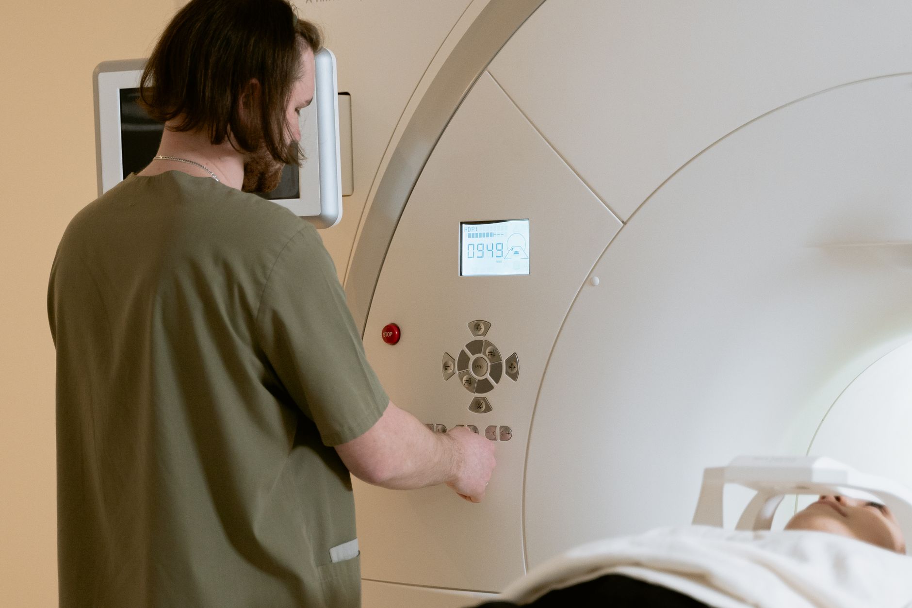
CT scans cannot directly detect Parkinson’s disease. A CT scan will not show the specific nerve cell damage, dopamine depletion, or Lewy bodies that define Parkinson’s disease at the tissue level. The diagnosis of Parkinson’s disease relies on clinical examination, medical history, and how a person responds to medication—not on imaging findings.
If a neurologist orders a CT scan for someone with tremor or movement problems, the scan is almost always meant to rule out other treatable conditions, not to confirm or exclude Parkinson’s. That said, CT scans play an important role in the diagnostic workup. A 65-year-old woman presenting with a year of progressive stiffness and slow movement might have Parkinson’s, but she could also have had a series of small strokes, a brain tumor, or normal pressure hydrocephalus. The CT scan can reveal these alternative diagnoses within minutes and prevent months of misdiagnosis and wrong treatment.
Table of Contents
- What Can a CT Scan Show in Someone with Parkinson’s Symptoms?
- CT Scans Versus MRI for Parkinson’s Diagnosis
- What Conditions CT Scans Can Rule Out
- The Role of CT Scanning in a Typical Parkinson’s Diagnostic Pathway
- Why CT Cannot Detect Early Parkinson’s Disease
- When Doctors Order CT for Suspected Parkinson’s
- Real-World Scenarios Where CT Changed Patient Management
What Can a CT Scan Show in Someone with Parkinson’s Symptoms?
A CT scan of the brain is a fast way to look for structural problems. It takes only a few minutes, uses a lower dose of radiation than a full body scan, and is widely available even in small hospitals. The CT will clearly show bleeding in the brain, large tumors, signs of previous strokes, hydrocephalus (fluid buildup in the ventricles), and significant brain shrinkage. If the CT is normal—meaning no bleeding, no tumor, no stroke, no hydrocephalus—it tells the neurologist that the symptoms are not caused by a sudden structural disaster.
However, a normal CT does not prove someone has Parkinson’s disease. Parkinson’s disease changes the brain at a microscopic level: dopamine neurons die, and protein deposits (Lewy bodies) form inside nerve cells. CT resolution is not fine enough to see these changes. An mri is better at seeing fine brain details, but even a normal MRI does not exclude Parkinson’s. The clinical examination and the patient’s response to levodopa (a dopamine replacement medication) remain the gold standard for diagnosis.
CT Scans Versus MRI for Parkinson’s Diagnosis
MRI provides much sharper images of soft brain tissue than CT does. If a neurologist suspects Parkinson’s but also wants to exclude small strokes or subtle brain changes, an MRI is the better choice. However, MRI takes 30 to 60 minutes, costs significantly more, and cannot be performed if someone has certain metal implants (like some pacemakers or cochlear implants). CT is faster, cheaper, and safer for these patients, making it useful as a first screening step.
A key limitation of both CT and MRI is that neither one can see the actual cell death and protein deposits of Parkinson’s disease. Both imaging methods can only detect secondary changes—swelling, shrinkage, or damage caused by something else. For this reason, many neurologists do not order any brain imaging at all for a straightforward case of Parkinson’s. They rely instead on the clinical picture and the medication trial. Imaging gets ordered when the presentation is atypical, the symptoms started very quickly, or the person is young and the diagnosis is uncertain.
What Conditions CT Scans Can Rule Out
A 58-year-old man came to his doctor with two months of increasing rigidity and trouble with fine motor movements in his left hand. His adult daughter had Parkinson’s, so he was worried. The neurologist ordered a CT scan not because she suspected Parkinson’s, but because the symptoms had appeared so rapidly. The CT revealed a meningioma—a usually benign brain tumor—pressing on the motor cortex. Surgery removed the tumor, and his symptoms resolved completely.
He did not have Parkinson’s at all. CT scans are particularly valuable at detecting stroke, bleeding, and subdural hematomas (bleeding between the brain and its outer covering). In older adults who fall and develop new movement problems, a CT scan can quickly determine whether a head injury caused bleeding that needs urgent surgery. CT can also show signs of vascular dementia—multiple small strokes—which can mimic Parkinson’s and cause movement slowing, stiffness, and gait problems. Finding evidence of multiple strokes changes the entire treatment strategy.
The Role of CT Scanning in a Typical Parkinson’s Diagnostic Pathway
Most people with Parkinson’s disease are diagnosed without any brain imaging. A neurologist sees a patient with resting tremor, stiffness, and slowed movement; the symptoms fit the clinical criteria; and a trial of levodopa produces a clear improvement. That is enough.
Imaging is not routine in uncomplicated cases because the CT and MRI findings will be normal, and the cost and time are not justified. However, if the symptom pattern is unusual—for example, very rapid symptom progression, symptoms starting in only one limb and not spreading, significant cognitive decline very early, or severe gait problems without much tremor—then imaging becomes more important. A CT scan at this stage serves as a safety net. It answers the question: “Is there something else going on here that I am missing?” This practical approach balances diagnostic accuracy with efficiency and cost.
Why CT Cannot Detect Early Parkinson’s Disease
One of the most important limitations of CT is that it cannot see Parkinson’s disease in its early stages, even though MRI technology has improved. In the earliest phases of Parkinson’s, there may be no visible brain shrinkage, no structural change, and no abnormality on any standard imaging. The dopamine neurons are dying, but the brain structure looks normal on imaging. This means that a person in the very early stages of Parkinson’s disease could have a completely normal CT scan—and also a completely normal MRI.
This is why some research centers use advanced imaging techniques like PET scans to look for dopamine deficiency in suspected early Parkinson’s disease. PET is more sensitive than CT or even standard MRI and can show dopamine loss in specific brain regions. However, PET is expensive, not widely available, and not part of routine diagnostic practice. For a patient with concerning symptoms but a normal CT, the next step is usually specialist evaluation by a neurologist and sometimes a trial of medication rather than more imaging.
When Doctors Order CT for Suspected Parkinson’s
A CT scan is most likely to be ordered when a patient first presents with symptoms and the clinical picture is not clear-cut. Primary care doctors often order a CT as a reassurance test—to exclude serious causes like tumor or stroke before referring to a neurologist. Emergency departments use CT to screen people who arrive with acute movement problems, dizziness, or falls.
In these settings, CT is a quick way to rule out immediate danger. Neurologists also order CT when evaluating atypical Parkinsonian syndromes—conditions that look like Parkinson’s but are caused by something else. Progressive supranuclear palsy, corticobasal degeneration, and multiple system atrophy all produce movement problems that can resemble Parkinson’s disease. A CT or MRI might reveal brain atrophy in a pattern or location that points toward one of these rare syndromes instead.
Real-World Scenarios Where CT Changed Patient Management
A 72-year-old woman developed progressive stiffness and a shuffling gait over eight months. Her family attributed it to old age, but her daughter insisted on a neurologist visit. The neurologist was concerned about Parkinson’s disease but also noticed that the woman had a history of high blood pressure and diabetes. A CT scan showed multiple old strokes in the deep white matter of her brain—classic vascular parkinsonism. She was started on blood pressure medication and a stroke-prevention medication rather than Parkinson’s drugs.
Her symptoms did not worsen, but they did not improve either, which is typical for vascular parkinsonism since the strokes cannot be reversed. Another patient, a 55-year-old man, presented with tremor that started in his right hand six months earlier. He had no family history of Parkinson’s disease and was highly functional. His CT scan was normal, his neurological exam was consistent with early Parkinson’s disease, and a trial of carbidopa-levodopa brought significant improvement. He proceeded with standard Parkinson’s care. In this straightforward case, the normal CT scan provided reassurance, and the diagnosis and treatment moved forward without delay.
- —