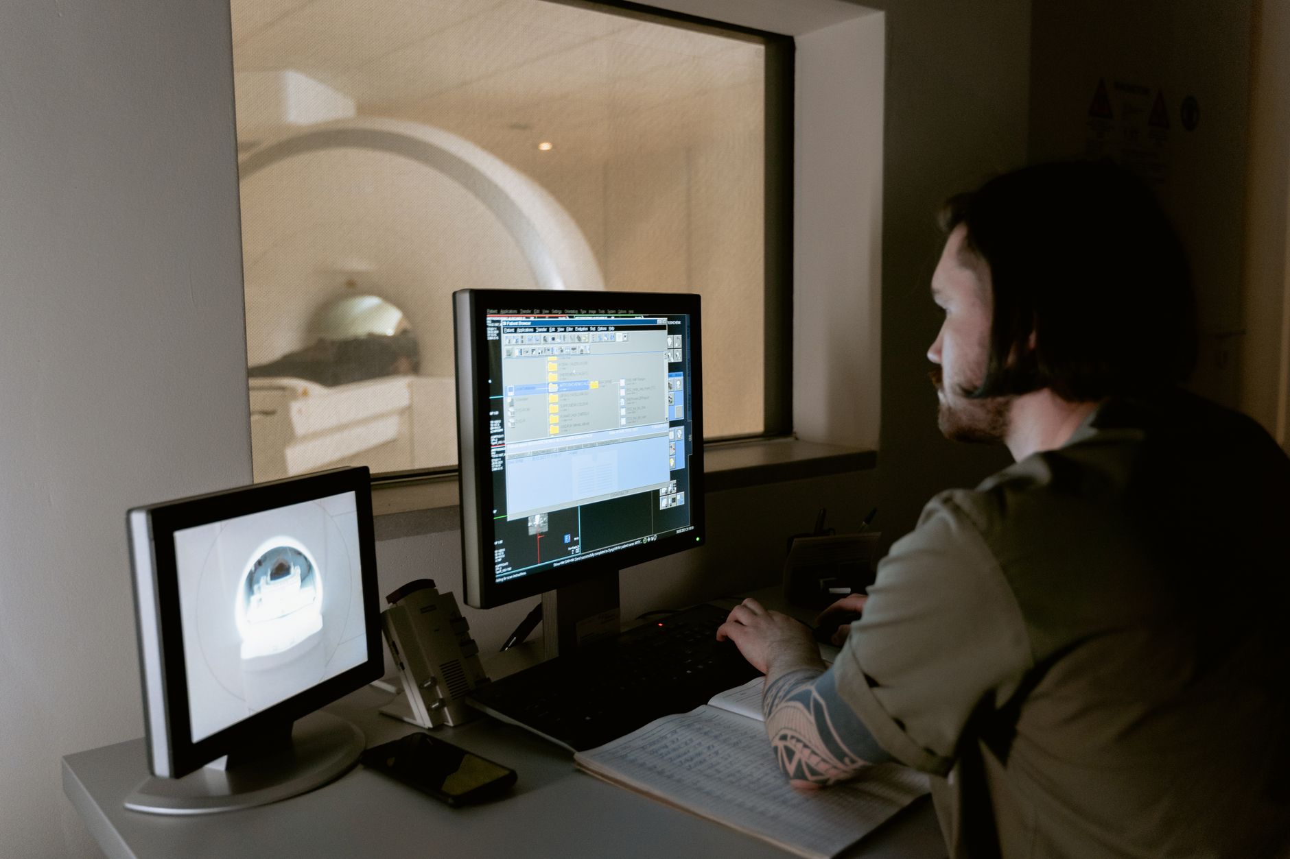
MRI cannot directly diagnose Parkinson’s disease, but it can reveal certain changes in the brain that may support a Parkinson’s diagnosis and, more importantly, rule out other conditions that cause similar symptoms. An MRI scan can show increased iron accumulation in the substantia nigra—a region of the brain central to Parkinson’s—along with changes in the size and shape of structures deep in the brain.
However, these findings alone are not specific enough to confirm Parkinson’s on their own, because people without Parkinson’s can have similar brain changes, and some people with Parkinson’s may show no obvious MRI abnormalities. When a neurologist suspects Parkinson’s disease, an MRI is often ordered not to prove someone has Parkinson’s, but to eliminate other diseases that can mimic Parkinson’s symptoms, such as normal-pressure hydrocephalus, multiple system atrophy, or vascular parkinsonism. In this role, MRI is a valuable safety tool that prevents misdiagnosis and ensures patients receive appropriate treatment.
Table of Contents
- What Does an MRI Show in Parkinson’s Disease Brains?
- Why MRI Cannot Diagnose Parkinson’s on Its Own
- The Real Role of MRI in Diagnosing Parkinson’s
- When Neurologists Order MRI for Suspected Parkinson’s
- Limitations and Common Pitfalls
- Other Imaging Tests That Play a Role in Parkinson’s Diagnosis
- What to Expect During an MRI Scan
- Frequently Asked Questions
What Does an MRI Show in Parkinson’s Disease Brains?
MRI scans can visualize several brain changes associated with Parkinson’s. The substantia nigra, a region roughly the size of a pea located deep in the midbrain, typically shows increased iron content in Parkinson’s patients—iron appears as a darker signal on certain MRI sequences. The putamen and globus pallidus, part of the brain’s motor control system, may also show changes in signal intensity or a loss of the normal distinction between these structures. Some patients display a characteristic pattern called the “swallow tail sign,” where the tail-like appearance of the substantia nigra is lost.
Despite these observable changes, researchers have found significant variation among Parkinson’s patients. One patient might show obvious iron accumulation while another shows very little, yet both may have identical symptoms. This variability is one reason why MRI findings cannot stand alone as a diagnostic test. Additionally, a small percentage of people with confirmed Parkinson’s disease show essentially normal MRI scans, making the absence of findings unreliable for ruling out the disease.
Why MRI Cannot Diagnose Parkinson’s on Its Own
The fundamental problem is that MRI shows brain structure and composition, but Parkinson’s disease is ultimately a disorder of brain chemistry and cellular loss that develops at a microscopic level. MRI can detect changes in iron, fluid, or the size of brain structures, but it cannot visualize the loss of dopamine-producing cells or the accumulation of alpha-synuclein protein—the hallmark pathological features of Parkinson’s. These crucial changes require examination of brain tissue under a microscope, which is only possible at autopsy.
Another critical limitation is specificity: brain changes visible on MRI in a Parkinson’s patient may also appear in people with other neurodegenerative diseases or even in healthy older adults. A radiologist looking at an MRI of someone over 70 might see age-related changes, iron accumulation, or subtle structural shifts that could suggest Parkinson’s, but these same findings could indicate normal aging, mild cognitive impairment, or an entirely different condition. This overlap makes it dangerous to rely on MRI alone, especially if the patient’s clinical history and examination findings don’t align with what the scan shows.
The Real Role of MRI in Diagnosing Parkinson’s
In clinical practice, MRI serves as a gate-keeping tool. When a person comes to a neurologist with symptoms like tremor, stiffness, and slow movement—symptoms that could be caused by Parkinson’s or by several other diseases—the doctor orders an MRI to look for findings that would point to an alternative diagnosis. If the MRI shows clear signs of a stroke, hydrocephalus (abnormal fluid buildup), a tumor, or multiple small vessel changes characteristic of vascular parkinsonism, the neurologist knows the symptoms are not due to Parkinson’s disease and can pursue a different treatment path.
For example, a patient presenting with slowness and rigidity undergoes an MRI that reveals moderate to severe enlargement of the brain’s ventricles (fluid-filled spaces) along with characteristic periventricular white matter changes. These findings, combined with the presence of gait imbalance and urinary incontinence, suggest normal-pressure hydrocephalus rather than Parkinson’s—a diagnosis that could be missed if the clinician relied only on symptoms. In this scenario, the MRI prevented a patient from being treated for a disease they do not have and instead directed them toward the correct diagnosis.
When Neurologists Order MRI for Suspected Parkinson’s
MRI is typically ordered early in the diagnostic workup of someone suspected of having Parkinson’s disease. The scan is especially important in patients presenting with “atypical” features that raise concern for a different condition—such as early cognitive decline, prominent vertical gaze difficulty (trouble looking up), or very rapid symptom progression. A person with a 15-year history of progressive tremor and slowness that fits the classic Parkinson’s picture might not require an MRI if the clinical diagnosis is clear, whereas someone with only 6 months of symptoms and additional warning signs absolutely should have one.
The tradeoff here is practical and financial. MRI is expensive, takes 30 to 60 minutes to complete, and requires the patient to hold still in a confined space—potentially difficult for someone with Parkinson’s tremor or rigidity. However, this cost and discomfort are justified by the diagnostic value it provides, particularly in ruling out reversible conditions like normal-pressure hydrocephalus, for which a specific treatment (ventriculoperitoneal shunt placement) exists. A mislabeled diagnosis that leads to years of inappropriate treatment is far more costly than one preventive MRI scan.
Limitations and Common Pitfalls
One of the most important limitations is that MRI findings can change with age and do not remain constant over time. A patient scanned at age 65 may show subtle iron changes, but a second MRI five years later at age 70 might show more extensive changes not because Parkinson’s has progressed, but because normal aging and iron accumulation continue regardless of disease. This makes it difficult for doctors to use serial MRI scans (multiple scans over time) to track Parkinson’s progression, even though such tracking is sometimes attempted.
Another pitfall occurs when radiologists or clinicians over-interpret MRI findings. Finding increased iron in the substantia nigra on MRI does not confirm Parkinson’s disease—it raises suspicion, but the final diagnosis still depends on clinical examination, the patient’s symptom history, and response to dopamine-replacement therapy. A patient might be labeled with Parkinson’s based primarily on an MRI report stating “findings consistent with Parkinson’s,” only to discover years later that the symptoms stem from a different condition or that the patient does not actually respond to standard Parkinson’s medications. The imaging serves a supporting role, never the primary role.
Other Imaging Tests That Play a Role in Parkinson’s Diagnosis
While MRI looks at brain structure, other imaging methods can provide complementary information. DAT (dopamine transporter) scans and FP-CIT SPECT imaging can detect loss of dopamine-producing cells in the striatum, offering more direct evidence of Parkinson’s pathology than MRI can provide.
However, these nuclear imaging tests are more expensive, involve radiation exposure, and are not routinely used for initial diagnosis in many clinical settings. PET imaging can also show changes in glucose metabolism and regional brain activity, but again, it is typically reserved for research or complex diagnostic cases. For most patients, the diagnostic pathway relies primarily on clinical assessment by a neurologist experienced in movement disorders, supported by MRI to rule out mimics and DAT scan or response to dopamine therapy to confirm Parkinson’s when the diagnosis remains uncertain.
What to Expect During an MRI Scan
If a neurologist orders an MRI as part of evaluating symptoms, the patient typically reports to a radiology department where they change into a hospital gown and remove all metal objects (jewelry, hearing aids, metal implants). The technician positions the patient on a sliding table and guides them into the MRI machine—a large cylindrical magnet about 60 inches long. The patient lies still for 30 to 60 minutes while the machine makes loud knocking and humming sounds as it creates detailed cross-sectional images of the brain.
The procedure itself is painless and involves no radiation, but it can be claustrophobic for some people, and the loud noise may be distressing. Patients with Parkinson’s disease who experience significant tremor, rigidity, or difficulty lying still sometimes require mild sedation to complete the scan. The radiologist reviews the images and generates a report within hours or days, which the ordering neurologist discusses with the patient during a follow-up visit.
Frequently Asked Questions
Can an MRI definitively prove I have Parkinson’s disease?
No. While an MRI can show brain changes associated with Parkinson’s, such as iron accumulation in the substantia nigra, these findings alone cannot diagnose the disease. Diagnosis requires clinical evaluation by a neurologist, including examination of symptoms and movement, combined with the patient’s response to Parkinson’s medications.
Will my neurologist order an MRI if I have Parkinson’s symptoms?
Likely yes, but not necessarily to confirm Parkinson’s. The MRI is ordered early in the diagnostic process to rule out other conditions that can mimic Parkinson’s symptoms, such as normal-pressure hydrocephalus, stroke, or brain tumors.
What if my MRI shows nothing abnormal—does that mean I don’t have Parkinson’s?
Not necessarily. Some people with confirmed Parkinson’s disease have relatively normal-appearing MRI scans. The brain changes visible on MRI vary widely among patients with the same diagnosis.
How long does an MRI scan take?
The scan itself typically lasts 30 to 60 minutes. The patient lies still inside the MRI machine while it captures detailed images of the brain. The procedure is painless and uses no radiation.
Are there any risks to having an MRI?
MRI is very safe for most people. The main concerns are discomfort from confinement (for people with claustrophobia) and difficulty holding still (relevant for patients with Parkinson’s tremor). Patients with certain metal implants or pacemakers may not be candidates for MRI; the technician will ask about this before the scan.
If MRI can’t diagnose Parkinson’s, what can?
Parkinson’s is diagnosed clinically by a neurologist based on examination findings (tremor at rest, rigidity, slowness of movement) and the patient’s response to levodopa (a dopamine-replacement medication). Additional tests like DAT scan or FP-CIT SPECT imaging can support the diagnosis in uncertain cases.