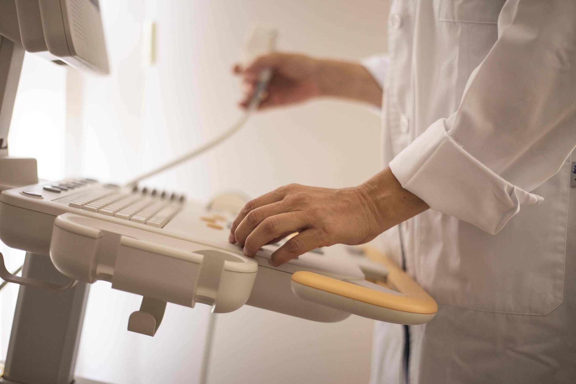
High-resolution ultrasound can detect a smaller vagus nerve cross-sectional area in some people with Parkinson’s disease, a finding often described as vagus nerve atrophy. Researchers are studying this change as a possible marker of autonomic nerve involvement, but the evidence is mixed, and vagus nerve ultrasound cannot currently diagnose Parkinson’s disease. For example, a neurologist might identify a reduced nerve measurement during a neck scan, yet still rely on symptoms, examination findings, medication response, and other testing to evaluate the patient.
The vagus nerve helps regulate automatic functions including heart rate, digestion, swallowing, and aspects of blood-pressure control. These functions can be disrupted in Parkinson’s disease, sometimes years before prominent tremor or stiffness develops. Ultrasound offers a relatively quick, noninvasive way to examine the nerve in the neck, but a small measurement does not reveal why the nerve is small or establish that it is functioning abnormally.
Medical information disclaimer: This article is for general educational purposes only and does not provide medical advice, diagnosis, or treatment. Always consult a physician or other qualified health professional about symptoms, medications, tests, or treatment decisions.
Table of Contents
- Can Ultrasound Detect Vagus Nerve Atrophy in Parkinson’s Disease?
- What Vagus Nerve Atrophy May Reveal About Parkinson’s Disease
- Vagus Nerve Changes and Autonomic Symptoms
- How Vagus Nerve Ultrasound Is Performed and Interpreted
- Conflicting Findings and Common Measurement Problems
- Implications for Parkinson’s Treatment and Medication
- Caregiving for Autonomic Symptoms Linked to Parkinson’s
- Frequently Asked Questions
Can Ultrasound Detect Vagus Nerve Atrophy in Parkinson’s Disease?
Ultrasound can visualize the cervical vagus nerve as it travels through the neck near the carotid artery and internal jugular vein. With a high-frequency probe, a trained examiner can outline the nerve and estimate its cross-sectional area. Some studies have found that this area is smaller on one or both sides in groups with Parkinson’s disease than in control groups. The result is a group-level association, not a dependable individual diagnostic test.
A person with Parkinson’s disease may have a vagus nerve measurement within the expected range, while someone without Parkinson’s may have a relatively small nerve. This overlap is similar to many imaging findings in neurology: a scan may support research into disease mechanisms without being specific enough to settle a diagnosis in the clinic. Measurements can also vary with probe position, image resolution, tracing technique, and the examiner’s experience. Even gentle pressure from the probe may alter the appearance of small structures in the neck. Studies therefore need standardized methods and blinded measurements before results from different medical centers can be compared confidently.
What Vagus Nerve Atrophy May Reveal About Parkinson’s Disease
The vagus nerve carries signals between the brainstem and organs in the chest and abdomen. It contains sensory, motor, and autonomic fibers, so structural changes could reflect broader involvement of the autonomic nervous system. This is relevant because constipation, delayed stomach emptying, abnormal sweating, and blood-pressure changes are common nonmotor features of Parkinson’s disease. Researchers have also investigated whether disease-related changes might travel between the gastrointestinal nervous system and the brain through vagal pathways.
This idea is connected to observations that abnormal alpha-synuclein can occur in nervous tissue outside the brain. However, finding a smaller cervical vagus nerve does not prove where Parkinson’s disease began or show that abnormal proteins traveled along that nerve. There is an important biological limitation: cross-sectional area is only a structural measurement. It cannot distinguish loss of nerve fibers from ordinary anatomical variation, aging, metabolic disease, inflammation, or technical error. It also does not directly measure nerve signaling, so “atrophy” should not automatically be interpreted as complete loss of vagal function.
Vagus Nerve Changes and Autonomic Symptoms
Autonomic symptoms can have a substantial effect on daily life. A person may become lightheaded after standing, feel full after a few bites, experience persistent constipation, or have unpredictable sweating. Because the vagus nerve participates in several automatic processes, researchers have examined whether smaller ultrasound measurements correspond with greater autonomic symptom burden. The relationship is not straightforward.
Orthostatic hypotension, for example, involves multiple parts of the autonomic nervous system and may also be worsened by dehydration, blood-pressure medicines, dopaminergic treatment, or prolonged inactivity. A small vagus nerve on ultrasound cannot identify which of these factors is responsible. Consider a person with Parkinson’s disease who develops nausea and delayed medication benefit after meals. Impaired stomach emptying may be contributing, but an ultrasound measurement of the neck portion of the vagus nerve would not confirm gastroparesis. The clinician may instead review meal timing, constipation, weight changes, and medication use, then consider gastrointestinal testing when appropriate.
How Vagus Nerve Ultrasound Is Performed and Interpreted
During the examination, the patient usually lies on their back while a clinician places an ultrasound probe along each side of the neck. The vagus nerve is located within the carotid sheath, generally between or behind major blood vessels. The examiner captures images and traces the nerve boundary to calculate its cross-sectional area. The procedure does not use ionizing radiation and is generally more accessible than specialized brain imaging.
It can also be repeated, which is useful in research following participants over time. The tradeoff is that ultrasound is highly operator-dependent: magnetic resonance imaging may provide more consistent anatomical context, while ultrasound offers portability and real-time imaging at a lower level of technical complexity. Patients should not change Parkinson’s medication or autonomic treatment because of an isolated ultrasound result. If a report mentions vagus nerve atrophy, useful questions include whether both sides were measured, whether the laboratory has reference values, and whether the finding changes any clinical decision. In many settings, it remains an investigational observation rather than an actionable diagnosis.
Conflicting Findings and Common Measurement Problems
Research findings have not been uniform. Some investigations report smaller vagus nerves in Parkinson’s disease, while others find little or no meaningful difference after accounting for variation among participants and measurement methods. Differences in disease duration, autonomic symptoms, body size, age, and coexisting conditions may partly explain the inconsistency. The nerve is small, and its ultrasound border can be difficult to separate from surrounding tissue.
A minor change in how the outline is traced can materially affect the calculated area. Comparing a measurement from one clinic with a reference range produced using different equipment or a different anatomical location may therefore be misleading. Another warning concerns diagnostic overreach. Vagus nerve atrophy is not established as a substitute for a neurological examination, and it does not distinguish Parkinson’s disease reliably from atypical parkinsonian disorders, peripheral neuropathies, or other causes of autonomic dysfunction. New fainting, repeated falls, severe swallowing trouble, unexplained weight loss, or persistent vomiting requires clinical assessment regardless of what an ultrasound shows.
Implications for Parkinson’s Treatment and Medication
Vagus nerve ultrasound does not currently determine which Parkinson’s medication a patient should receive. Levodopa and other treatments are selected according to movement symptoms, daily functioning, side effects, age, and associated medical problems—not the measured size of the cervical vagus nerve.
The scan may still contribute to research on symptom patterns and treatment response. For example, investigators could compare nerve measurements with constipation severity or blood-pressure changes over time. In routine care, however, a medication dose that seems slow or unreliable is more likely to prompt a review of meal protein, gastric emptying, constipation, and dosing schedules than a vagus nerve scan.
Caregiving for Autonomic Symptoms Linked to Parkinson’s
Caregivers can help document symptoms that may otherwise appear unrelated. A practical record might note when dizziness occurs, whether it follows standing or meals, bowel-movement frequency, fluid intake, falls, swallowing problems, and the timing of Parkinson’s medication. Blood-pressure readings taken while lying down and again after standing may also help a clinician investigate orthostatic symptoms when performed according to medical instructions.
Safety measures should match the symptom rather than an ultrasound finding. Someone who becomes dizzy on standing may need to rise slowly and pause before walking, while a person who coughs during meals may need a swallowing assessment. Sudden fainting, chest pain, signs of dehydration, or an inability to swallow medication warrants prompt medical attention.
Frequently Asked Questions
Can vagus nerve ultrasound diagnose Parkinson’s disease?
No. Parkinson’s disease remains a clinical diagnosis based primarily on medical history and neurological examination. Ultrasound findings may support research, but they are not sufficiently specific or consistent for independent diagnosis.
Is vagus nerve ultrasound painful?
The examination is generally painless and uses a probe placed on the skin of the neck. Patients may feel mild pressure from the probe and ultrasound gel.
Does a smaller vagus nerve explain constipation in Parkinson’s disease?
Not by itself. Constipation may involve slowed intestinal movement, reduced activity, diet, fluid intake, medications, and changes across several parts of the nervous system. Ultrasound cannot identify the cause in an individual patient.
Can vagus nerve atrophy be reversed?
Research has not established that an ultrasound-measured reduction can be reversed or that increasing nerve size would improve Parkinson’s symptoms. Treatment should focus on identified symptoms and clinically supported interventions.
Is vagus nerve stimulation a treatment for Parkinson’s disease?
Vagus nerve stimulation has established uses for certain other neurological or psychiatric conditions, but it is not a standard treatment for Parkinson’s disease. Experimental stimulation approaches should not be confused with diagnostic ultrasound.